 Movie
Movie Controller

[English] 日本語
 Yorodumi
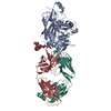
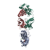
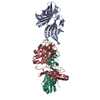
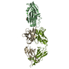
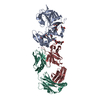
- PDB-7coe: Crystal structure of Receptor binding domain of MERS-CoV and KNIH... -
+ Open data

- Basic information
| Entry | Database: PDB / ID: 7coe | ||||||
|---|---|---|---|---|---|---|---|
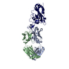
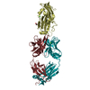
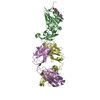
| Title | Crystal structure of Receptor binding domain of MERS-CoV and KNIH90-F1 Fab complex | ||||||
 Components Components |
| ||||||
 Keywords Keywords | IMMUNE SYSTEM / RBD / Fab / complex | ||||||
| Function / homology |  Function and homology information Function and homology informationhost cell endoplasmic reticulum-Golgi intermediate compartment membrane / receptor-mediated virion attachment to host cell / endocytosis involved in viral entry into host cell / fusion of virus membrane with host plasma membrane / fusion of virus membrane with host endosome membrane / viral envelope / host cell plasma membrane / virion membrane / membrane Similarity search - Function | ||||||
| Biological species |  Homo sapiens (human) Homo sapiens (human) 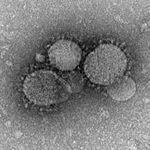 Middle East respiratory syndrome-related coronavirus Middle East respiratory syndrome-related coronavirus | ||||||
| Method |  X-RAY DIFFRACTION / SYNCHROTRON / MOLECULAR REPLACEMENT / Resolution: 2.05 Å X-RAY DIFFRACTION / SYNCHROTRON / MOLECULAR REPLACEMENT / Resolution: 2.05 Å | ||||||
 Authors Authors | Lee, J.Y. / Song, J.Y. / Lee, H.S. / Hong, E. / Jang, T.H. | ||||||
 Citation Citation | Journal: Sci Rep / Year: 2022 Title: The structure of a novel antibody against the spike protein inhibits Middle East respiratory syndrome coronavirus infections. Authors: Jang, T.H. / Park, W.J. / Lee, H. / Woo, H.M. / Lee, S.Y. / Kim, K.C. / Kim, S.S. / Hong, E. / Song, J. / Lee, J.Y. | ||||||
| History |
|
- Structure visualization
| Structure viewer | Molecule: MolmilJmol/JSmol |
|---|
- Downloads & links
-Download
| PDBx/mmCIF format | 7coe.cif.gz | 567.6 KB | Display | PDBx/mmCIF format |
|---|---|---|---|---|
| PDB format | pdb7coe.ent.gz | 445.6 KB | Display | PDB format |
| PDBx/mmJSON format | 7coe.json.gz | Tree view | PDBx/mmJSON format | |
| Others |  Other downloads Other downloads |
-Validation report
| Arichive directory | https://data.pdbj.org/pub/pdb/validation_reports/co/7coeftp://data.pdbj.org/pub/pdb/validation_reports/co/7coe | HTTPS FTP |
|---|
-Related structure data
| Related structure data |  4zs6S S: Starting model for refinement |
|---|---|
| Similar structure data |

-Links
 PDBj
PDBj

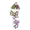
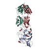
- Assembly
| Deposited unit | 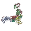
| ||||||||||||
|---|---|---|---|---|---|---|---|---|---|---|---|---|---|
| 1 | 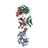
| ||||||||||||
| 2 | 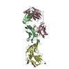
| ||||||||||||
| Unit cell |
|
-Components
-Antibody , 2 types, 4 molecules HBLC
| #1: Antibody | Mass: 25216.145 Da / Num. of mol.: 2 Source method: isolated from a genetically manipulated source Source: (gene. exp.) Homo sapiens (human) / Production host: Homo sapiens (human)#2: Antibody | Mass: 23259.771 Da / Num. of mol.: 2 Source method: isolated from a genetically manipulated source Source: (gene. exp.) Homo sapiens (human) / Production host: Homo sapiens (human) |
|---|
-Protein / Sugars , 2 types, 6 molecules AD
| #3: Protein | Mass: 24429.520 Da / Num. of mol.: 2 Source method: isolated from a genetically manipulated source Source: (gene. exp.) Middle East respiratory syndrome-related coronavirusProduction host:   Spodoptera frugiperda (fall armyworm) / References: UniProt: W6A0E0 Spodoptera frugiperda (fall armyworm) / References: UniProt: W6A0E0#5: Sugar | ChemComp-NAG /  Type: D-saccharide, beta linking / Mass: 221.208 Da / Num. of mol.: 4 Type: D-saccharide, beta linking / Mass: 221.208 Da / Num. of mol.: 4Source method: isolated from a genetically manipulated source Formula: C8H15NO6 / Feature type: SUBJECT OF INVESTIGATION |
|---|
-Non-polymers , 2 types, 999 molecules 

| #4: Chemical | ChemComp-EDO /  Mass: 62.068 Da / Num. of mol.: 29 / Source method: obtained synthetically / Formula: C2H6O2 Mass: 62.068 Da / Num. of mol.: 29 / Source method: obtained synthetically / Formula: C2H6O2#6: Water | ChemComp-HOH / | Mass: 18.015 Da / Num. of mol.: 970 / Source method: isolated from a natural source / Formula: H2O |
|---|
-Details
| Has ligand of interest | Y |
|---|---|
| Has protein modification | Y |
-Experimental details
-Experiment
| Experiment | Method: X-RAY DIFFRACTION / Number of used crystals: 1 |
|---|
- Sample preparation
| Crystal | Density Matthews: 2.92 Å3/Da / Density % sol: 57.9 % |
|---|---|
| Crystal grow | Temperature: 291 K / Method: vapor diffusion, hanging drop / pH: 6.3 / Details: 0.1M MES monohydrate pH 6.3, 15%(w/v) PEG 20000 |
-Data collection
| Diffraction | Mean temperature: 100 K / Serial crystal experiment: N |
|---|---|
| Diffraction source | Source: SYNCHROTRON / Site: PAL/PLS  / Beamline: 5C (4A) / Wavelength: 0.9793 Å / Beamline: 5C (4A) / Wavelength: 0.9793 Å |
| Detector | Type: DECTRIS EIGER X 9M / Detector: PIXEL / Date: Jun 20, 2019 |
| Radiation | Protocol: SINGLE WAVELENGTH / Monochromatic (M) / Laue (L): M / Scattering type: x-ray |
| Radiation wavelength | Wavelength: 0.9793 Å / Relative weight: 1 |
| Reflection | Resolution: 2.05→44.95 Å / Num. obs: 103522 / % possible obs: 98.95 % / Redundancy: 7 % / CC1/2: 0.956 / Net I/σ(I): 14.97 |
| Reflection shell | Resolution: 2.05→2.123 Å / Num. unique obs: 9762 / CC1/2: 0.896 |
- Processing
| Software |
| ||||||||||||||||||||||||
|---|---|---|---|---|---|---|---|---|---|---|---|---|---|---|---|---|---|---|---|---|---|---|---|---|---|
| Refinement | Method to determine structure: MOLECULAR REPLACEMENT Starting model: 4ZS6 Resolution: 2.05→44.95 Å / Cross valid method: FREE R-VALUE Stereochemistry target values: GeoStd + Monomer Library + CDL v1.2
| ||||||||||||||||||||||||
| Displacement parameters | Biso mean: 38.45 Å2 | ||||||||||||||||||||||||
| Refinement step | Cycle: LAST / Resolution: 2.05→44.95 Å
| ||||||||||||||||||||||||
| Refine LS restraints |
|
