 Movie
Movie Controller

[English] 日本語
 Yorodumi
- PDB-7vso: Serial Femtosecond Crystallography (SFX) of Ground State Bacterio... -
+ Open data

- Basic information
| Entry | Database: PDB / ID: 7vso | ||||||
|---|---|---|---|---|---|---|---|
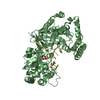
| Title | Serial Femtosecond Crystallography (SFX) of Ground State Bacteriorhodopsin Crystallized from Bicelles in Complex with HAD16 Determined Using 7-keV X-ray Free Electron Laser (XFEL) at SACLA | ||||||
 Components Components | Bacteriorhodopsin | ||||||
 Keywords Keywords | MEMBRANE PROTEIN / proton pump / transport protein / retinal / proton transport | ||||||
| Function / homology |  Function and homology information Function and homology informationlight-driven active monoatomic ion transmembrane transporter activity / monoatomic ion channel activity / photoreceptor activity / phototransduction / proton transmembrane transport / plasma membrane Similarity search - Function | ||||||
| Biological species |  Halobacterium salinarum (Halophile) Halobacterium salinarum (Halophile) | ||||||
| Method |  X-RAY DIFFRACTION / FREE ELECTRON LASER / MOLECULAR REPLACEMENT / Resolution: 2.35 Å X-RAY DIFFRACTION / FREE ELECTRON LASER / MOLECULAR REPLACEMENT / Resolution: 2.35 Å | ||||||
 Authors Authors | Mizohata, E. / Nakane, T. / Hanashima, S. | ||||||
| Funding support |  Japan, 1items Japan, 1items
| ||||||
 Citation Citation | Journal: Membranes (Basel) / Year: 2021 Title: Heavy Atom Detergent/Lipid Combined X-ray Crystallography for Elucidating the Structure-Function Relationships of Membrane Proteins. Authors: Hanashima, S. / Nakane, T. / Mizohata, E. | ||||||
| History |
|
- Structure visualization
| Structure viewer | Molecule: MolmilJmol/JSmol |
|---|
- Downloads & links
-Download
| PDBx/mmCIF format | 7vso.cif.gz | 71.6 KB | Display | PDBx/mmCIF format |
|---|---|---|---|---|
| PDB format | pdb7vso.ent.gz | 49.2 KB | Display | PDB format |
| PDBx/mmJSON format | 7vso.json.gz | Tree view | PDBx/mmJSON format | |
| Others |  Other downloads Other downloads |
-Validation report
| Summary document | 7vso_validation.pdf.gz | 3.7 MB | Display | wwPDB validaton report |
|---|---|---|---|---|
| Full document | 7vso_full_validation.pdf.gz | 3.8 MB | Display | |
| Data in XML | 7vso_validation.xml.gz | 14.4 KB | Display | |
| Data in CIF | 7vso_validation.cif.gz | 18.2 KB | Display | |
| Arichive directory | https://data.pdbj.org/pub/pdb/validation_reports/vs/7vsoftp://data.pdbj.org/pub/pdb/validation_reports/vs/7vso | HTTPS FTP |
-Related structure data
| Related structure data |  5b35S S: Starting model for refinement |
|---|---|
| Similar structure data | |
| Experimental dataset #1 | Data reference: 10.11577/1834012 / Data set type: diffraction image data |


-Links
 PDBj
PDBj


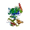
- Assembly
| Deposited unit | 
| ||||||||
|---|---|---|---|---|---|---|---|---|---|
| 1 | 
| ||||||||
| Unit cell |
| ||||||||
| Components on special symmetry positions |
|
-Components
-Protein , 1 types, 1 molecules A
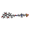
| #1: Protein | Mass: 26929.500 Da / Num. of mol.: 1 / Source method: isolated from a natural source / Source: (natural) Halobacterium salinarum (Halophile) / Strain: R1M1 / References: UniProt: P02945 |
|---|
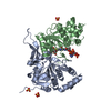
-Non-polymers , 11 types, 52 molecules 


| #2: Chemical | ChemComp-RET /  Mass: 284.436 Da / Num. of mol.: 1 / Source method: obtained synthetically / Formula: C20H28O / Feature type: SUBJECT OF INVESTIGATION Mass: 284.436 Da / Num. of mol.: 1 / Source method: obtained synthetically / Formula: C20H28O / Feature type: SUBJECT OF INVESTIGATION | ||||||||||||
|---|---|---|---|---|---|---|---|---|---|---|---|---|---|
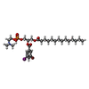
| #3: Chemical | ChemComp-7YH /  Mass: 777.483 Da / Num. of mol.: 1 / Source method: obtained synthetically / Formula: C29H49BrINO8P / Feature type: SUBJECT OF INVESTIGATION Mass: 777.483 Da / Num. of mol.: 1 / Source method: obtained synthetically / Formula: C29H49BrINO8P / Feature type: SUBJECT OF INVESTIGATION | ||||||||||||
| #4: Chemical | ChemComp-IOD /  Mass: 126.904 Da / Num. of mol.: 1 / Source method: obtained synthetically / Formula: I / Feature type: SUBJECT OF INVESTIGATION Mass: 126.904 Da / Num. of mol.: 1 / Source method: obtained synthetically / Formula: I / Feature type: SUBJECT OF INVESTIGATION | ||||||||||||
| #5: Chemical | ChemComp-CPS /  Mass: 614.877 Da / Num. of mol.: 1 / Source method: obtained synthetically / Formula: C32H58N2O7S / Feature type: SUBJECT OF INVESTIGATION / Comment: detergent*YM Mass: 614.877 Da / Num. of mol.: 1 / Source method: obtained synthetically / Formula: C32H58N2O7S / Feature type: SUBJECT OF INVESTIGATION / Comment: detergent*YM | ||||||||||||
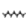
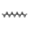
| #6: Chemical |  Mass: 114.229 Da / Num. of mol.: 2 / Source method: obtained synthetically / Formula: C8H18 / Feature type: SUBJECT OF INVESTIGATION Mass: 114.229 Da / Num. of mol.: 2 / Source method: obtained synthetically / Formula: C8H18 / Feature type: SUBJECT OF INVESTIGATION#7: Chemical | ChemComp-HP6 /  Mass: 100.202 Da / Num. of mol.: 5 / Source method: obtained synthetically / Formula: C7H16 / Feature type: SUBJECT OF INVESTIGATION Mass: 100.202 Da / Num. of mol.: 5 / Source method: obtained synthetically / Formula: C7H16 / Feature type: SUBJECT OF INVESTIGATION#8: Chemical |  Mass: 170.335 Da / Num. of mol.: 3 / Source method: obtained synthetically / Formula: C12H26 / Feature type: SUBJECT OF INVESTIGATION Mass: 170.335 Da / Num. of mol.: 3 / Source method: obtained synthetically / Formula: C12H26 / Feature type: SUBJECT OF INVESTIGATION#9: Chemical |  Mass: 226.441 Da / Num. of mol.: 3 / Source method: obtained synthetically / Formula: C16H34 / Feature type: SUBJECT OF INVESTIGATION Mass: 226.441 Da / Num. of mol.: 3 / Source method: obtained synthetically / Formula: C16H34 / Feature type: SUBJECT OF INVESTIGATION#10: Chemical |  Mass: 142.282 Da / Num. of mol.: 2 / Source method: obtained synthetically / Formula: C10H22 / Feature type: SUBJECT OF INVESTIGATION Mass: 142.282 Da / Num. of mol.: 2 / Source method: obtained synthetically / Formula: C10H22 / Feature type: SUBJECT OF INVESTIGATION#11: Chemical | ChemComp-DD9 / |  Mass: 128.255 Da / Num. of mol.: 1 / Source method: obtained synthetically / Formula: C9H20 / Feature type: SUBJECT OF INVESTIGATION Mass: 128.255 Da / Num. of mol.: 1 / Source method: obtained synthetically / Formula: C9H20 / Feature type: SUBJECT OF INVESTIGATION#12: Water | ChemComp-HOH / | Mass: 18.015 Da / Num. of mol.: 32 / Source method: isolated from a natural source / Formula: H2O |
-Details
| Has ligand of interest | Y |
|---|
-Experimental details
-Experiment
| Experiment | Method: X-RAY DIFFRACTION / Number of used crystals: 1 |
|---|
- Sample preparation
| Crystal | Density Matthews: 2.84 Å3/Da / Density % sol: 56.73 % |
|---|---|
| Crystal grow | Temperature: 293 K / Method: batch mode Details: 25%(W/V) DMPC/CHAPSO BICELLES, 3.2 M REMARK 280 NAH2PO4, 3.5%(W/V) TRIETHYLENE GLYCOL, 180 MM 1,6-HEXANEDIOL, 4 MM HAD16 |
-Data collection
| Diffraction | Mean temperature: 293 K / Serial crystal experiment: N |
|---|---|
| Diffraction source | Source: FREE ELECTRON LASER / Site: SACLA / Beamline: BL3 / Wavelength: 1.771 Å |
| Detector | Type: MPCCD / Detector: CCD / Date: Nov 22, 2015 |
| Radiation | Protocol: SINGLE WAVELENGTH / Monochromatic (M) / Laue (L): M / Scattering type: x-ray |
| Radiation wavelength | Wavelength: 1.771 Å / Relative weight: 1 |
| Reflection | Resolution: 2.35→42.9 Å / Num. obs: 13211 / % possible obs: 100 % / Redundancy: 206.2 % / CC1/2: 0.991 / Net I/σ(I): 7.34 |
| Reflection shell | Resolution: 2.35→2.43 Å / Num. unique obs: 897 / CC1/2: 0.488 |
- Processing
| Software |
| ||||||||||||||||||||||||||||||||||||||||||||||||||||||||||||||||||||||||||||||||||||||||||||||||||||||||||||||||||||||||||||||||||||||||||||||||||||||||||||||||||||||||||||||||||||||
|---|---|---|---|---|---|---|---|---|---|---|---|---|---|---|---|---|---|---|---|---|---|---|---|---|---|---|---|---|---|---|---|---|---|---|---|---|---|---|---|---|---|---|---|---|---|---|---|---|---|---|---|---|---|---|---|---|---|---|---|---|---|---|---|---|---|---|---|---|---|---|---|---|---|---|---|---|---|---|---|---|---|---|---|---|---|---|---|---|---|---|---|---|---|---|---|---|---|---|---|---|---|---|---|---|---|---|---|---|---|---|---|---|---|---|---|---|---|---|---|---|---|---|---|---|---|---|---|---|---|---|---|---|---|---|---|---|---|---|---|---|---|---|---|---|---|---|---|---|---|---|---|---|---|---|---|---|---|---|---|---|---|---|---|---|---|---|---|---|---|---|---|---|---|---|---|---|---|---|---|---|---|---|---|
| Refinement | Method to determine structure: MOLECULAR REPLACEMENT Starting model: 5B35 Resolution: 2.35→42.9 Å / Cor.coef. Fo:Fc: 0.964 / Cor.coef. Fo:Fc free: 0.947 / SU B: 7.393 / SU ML: 0.164 / Cross valid method: THROUGHOUT / ESU R: 0.262 / ESU R Free: 0.204 / Stereochemistry target values: MAXIMUM LIKELIHOOD / Details: HYDROGENS HAVE BEEN USED IF PRESENT IN THE INPUT
| ||||||||||||||||||||||||||||||||||||||||||||||||||||||||||||||||||||||||||||||||||||||||||||||||||||||||||||||||||||||||||||||||||||||||||||||||||||||||||||||||||||||||||||||||||||||
| Solvent computation | Ion probe radii: 0.8 Å / Shrinkage radii: 0.8 Å / VDW probe radii: 1.2 Å / Solvent model: BABINET MODEL WITH MASK | ||||||||||||||||||||||||||||||||||||||||||||||||||||||||||||||||||||||||||||||||||||||||||||||||||||||||||||||||||||||||||||||||||||||||||||||||||||||||||||||||||||||||||||||||||||||
| Displacement parameters | Biso mean: 79.264 Å2
| ||||||||||||||||||||||||||||||||||||||||||||||||||||||||||||||||||||||||||||||||||||||||||||||||||||||||||||||||||||||||||||||||||||||||||||||||||||||||||||||||||||||||||||||||||||||
| Refinement step | Cycle: 1 / Resolution: 2.35→42.9 Å
| ||||||||||||||||||||||||||||||||||||||||||||||||||||||||||||||||||||||||||||||||||||||||||||||||||||||||||||||||||||||||||||||||||||||||||||||||||||||||||||||||||||||||||||||||||||||
| Refine LS restraints |
|