 Movie
Movie Controller


+ Open data

- Basic information
| Entry | Database: PDB / ID: 7v6l | ||||||
|---|---|---|---|---|---|---|---|
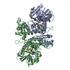
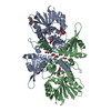
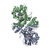
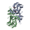
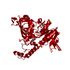
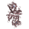
| Title | LcCOMT in complex with SAH | ||||||
 Components Components | LcCOMT | ||||||
 Keywords Keywords | TRANSFERASE / SAM / complex | ||||||
| Function / homology | S-ADENOSYL-L-HOMOCYSTEINE Function and homology information Function and homology information | ||||||
| Biological species |  Ligusticum chuanxiong (chuang-xiong) Ligusticum chuanxiong (chuang-xiong) | ||||||
| Method |  X-RAY DIFFRACTION / SYNCHROTRON / MOLECULAR REPLACEMENT / Resolution: 1.948 Å X-RAY DIFFRACTION / SYNCHROTRON / MOLECULAR REPLACEMENT / Resolution: 1.948 Å | ||||||
 Authors Authors | Yu, Y. / CHen, Q. | ||||||
| Funding support | 1items
| ||||||
 Citation Citation | Journal: Int.J.Biol.Macromol. / Year: 2022 Title: Structure basis of the caffeic acid O-methyltransferase from Ligusiticum chuanxiong to understand its selective mechanism. Authors: Song, S. / Chen, A. / Zhu, J. / Yan, Z. / An, Q. / Zhou, J. / Liao, H. / Yu, Y. | ||||||
| History |
|
- Structure visualization
| Structure viewer | Molecule: MolmilJmol/JSmol |
|---|
- Downloads & links
-Download
| PDBx/mmCIF format | 7v6l.cif.gz | 265.4 KB | Display | PDBx/mmCIF format |
|---|---|---|---|---|
| PDB format | pdb7v6l.ent.gz | 215.1 KB | Display | PDB format |
| PDBx/mmJSON format | 7v6l.json.gz | Tree view | PDBx/mmJSON format | |
| Others |  Other downloads Other downloads |
-Validation report
| Arichive directory | https://data.pdbj.org/pub/pdb/validation_reports/v6/7v6lftp://data.pdbj.org/pub/pdb/validation_reports/v6/7v6l | HTTPS FTP |
|---|
-Related structure data
| Related structure data | 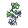 7v6jC 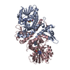 3reoS S: Starting model for refinement C: citing same article ( |
|---|---|
| Similar structure data |


-Links
 PDBj
PDBj- Assembly
| Deposited unit | 
| ||||||||
|---|---|---|---|---|---|---|---|---|---|
| 1 |
| ||||||||
| Unit cell |
|
-Components
| #1: Protein | Mass: 39954.176 Da / Num. of mol.: 2 Source method: isolated from a genetically manipulated source Source: (gene. exp.) Ligusticum chuanxiong (chuang-xiong) / Production host:  #2: Chemical |   Type: L-peptide linking / Mass: 384.411 Da / Num. of mol.: 2 / Source method: obtained synthetically / Formula: C14H20N6O5S Type: L-peptide linking / Mass: 384.411 Da / Num. of mol.: 2 / Source method: obtained synthetically / Formula: C14H20N6O5S#3: Water | ChemComp-HOH / |  Mass: 18.015 Da / Num. of mol.: 329 / Source method: isolated from a natural source / Formula: H2O Mass: 18.015 Da / Num. of mol.: 329 / Source method: isolated from a natural source / Formula: H2OHas ligand of interest | N | |
|---|
-Experimental details
-Experiment
| Experiment | Method: X-RAY DIFFRACTION / Number of used crystals: 1 |
|---|
- Sample preparation
| Crystal | Density Matthews: 2.26 Å3/Da / Density % sol: 45.56 % |
|---|---|
| Crystal grow | Temperature: 293 K / Method: vapor diffusion, hanging drop / pH: 6.5 Details: 0.2 M Ammonium acetate, 0.1 M BIS-TRIS pH 6.5, 25% w/v Polyethylene glycol 3350 |
-Data collection
| Diffraction | Mean temperature: 100 K / Serial crystal experiment: N |
|---|---|
| Diffraction source | Source: SYNCHROTRON / Site: SSRF  / Beamline: BL19U1 / Wavelength: 0.97774 Å / Beamline: BL19U1 / Wavelength: 0.97774 Å |
| Detector | Type: DECTRIS PILATUS 6M / Detector: PIXEL / Date: May 10, 2018 |
| Radiation | Protocol: SINGLE WAVELENGTH / Monochromatic (M) / Laue (L): M / Scattering type: x-ray |
| Radiation wavelength | Wavelength: 0.97774 Å / Relative weight: 1 |
| Reflection | Resolution: 1.948→50 Å / Num. obs: 51456 / % possible obs: 98.7 % / Redundancy: 6.6 % / Rrim(I) all: 0.129 / Net I/σ(I): 14 |
| Reflection shell | Resolution: 1.948→2 Å / Num. unique obs: 3108 / CC1/2: 0.939 |
- Processing
| Software |
| ||||||||||||||||||||||||||||||||||||||||||||||||||||||||||||||||||||||||||||||||||||||||||||||||||||||
|---|---|---|---|---|---|---|---|---|---|---|---|---|---|---|---|---|---|---|---|---|---|---|---|---|---|---|---|---|---|---|---|---|---|---|---|---|---|---|---|---|---|---|---|---|---|---|---|---|---|---|---|---|---|---|---|---|---|---|---|---|---|---|---|---|---|---|---|---|---|---|---|---|---|---|---|---|---|---|---|---|---|---|---|---|---|---|---|---|---|---|---|---|---|---|---|---|---|---|---|---|---|---|---|
| Refinement | Method to determine structure: MOLECULAR REPLACEMENT Starting model: 3reo Resolution: 1.948→44.053 Å / SU ML: 0.21 / Cross valid method: THROUGHOUT / σ(F): 1.35 / Phase error: 23.64 / Stereochemistry target values: ML
| ||||||||||||||||||||||||||||||||||||||||||||||||||||||||||||||||||||||||||||||||||||||||||||||||||||||
| Solvent computation | Shrinkage radii: 0.9 Å / VDW probe radii: 1.11 Å / Solvent model: FLAT BULK SOLVENT MODEL | ||||||||||||||||||||||||||||||||||||||||||||||||||||||||||||||||||||||||||||||||||||||||||||||||||||||
| Displacement parameters | Biso max: 109.42 Å2 / Biso mean: 27.3956 Å2 / Biso min: 2.9 Å2 | ||||||||||||||||||||||||||||||||||||||||||||||||||||||||||||||||||||||||||||||||||||||||||||||||||||||
| Refinement step | Cycle: final / Resolution: 1.948→44.053 Å
| ||||||||||||||||||||||||||||||||||||||||||||||||||||||||||||||||||||||||||||||||||||||||||||||||||||||
| LS refinement shell | Refine-ID: X-RAY DIFFRACTION / Rfactor Rfree error: 0
|
