 Movie
Movie Controller

[English] 日本語
 Yorodumi
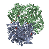
- PDB-2gz3: Structure of Aspartate Semialdehyde Dehydrogenase (ASADH) from St... -
+ Open data

- Basic information
| Entry | Database: PDB / ID: 2gz3 | ||||||
|---|---|---|---|---|---|---|---|
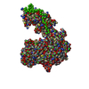
| Title | Structure of Aspartate Semialdehyde Dehydrogenase (ASADH) from Streptococcus pneumoniae complexed with NADP and aspartate-semialdehyde | ||||||
 Components Components | Aspartate beta-semialdehyde dehydrogenase | ||||||
 Keywords Keywords | OXIDOREDUCTASE / dehydrogenase / aspartate pathway | ||||||
| Function / homology |  Function and homology information Function and homology informationaspartate-semialdehyde dehydrogenase / aspartate-semialdehyde dehydrogenase activity / 'de novo' L-methionine biosynthetic process / L-threonine biosynthetic process / : / : / : / NAD binding / NADP binding / protein dimerization activity Similarity search - Function | ||||||
| Biological species |   Streptococcus pneumoniae (bacteria) Streptococcus pneumoniae (bacteria) | ||||||
| Method |  X-RAY DIFFRACTION / MOLECULAR REPLACEMENT / Resolution: 2.1 Å X-RAY DIFFRACTION / MOLECULAR REPLACEMENT / Resolution: 2.1 Å | ||||||
 Authors Authors | Faehnle, C.R. / Le Coq, J. / Liu, X. / Viola, R.E. | ||||||
 Citation Citation | Journal: J.Biol.Chem. / Year: 2006 Title: Examination of key intermediates in the catalytic cycle of aspartate-beta-semialdehyde dehydrogenase from a gram-positive infectious bacteria. Authors: Faehnle, C.R. / Le Coq, J. / Liu, X. / Viola, R.E. | ||||||
| History |
|
- Structure visualization
| Structure viewer | Molecule: MolmilJmol/JSmol |
|---|
- Downloads & links
-Download
| PDBx/mmCIF format | 2gz3.cif.gz | 535.1 KB | Display | PDBx/mmCIF format |
|---|---|---|---|---|
| PDB format | pdb2gz3.ent.gz | 442.6 KB | Display | PDB format |
| PDBx/mmJSON format | 2gz3.json.gz | Tree view | PDBx/mmJSON format | |
| Others |  Other downloads Other downloads |
-Validation report
| Arichive directory | https://data.pdbj.org/pub/pdb/validation_reports/gz/2gz3ftp://data.pdbj.org/pub/pdb/validation_reports/gz/2gz3 | HTTPS FTP |
|---|
-Related structure data
| Related structure data | 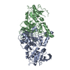 2gyyC 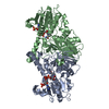 2gz1SC  2gz2C C: citing same article ( S: Starting model for refinement |
|---|---|
| Similar structure data |


-Links
 PDBj
PDBj- Assembly
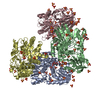
| Deposited unit | 
| ||||||||
|---|---|---|---|---|---|---|---|---|---|
| 1 |
| ||||||||
| 2 | 
| ||||||||
| 3 | 
| ||||||||
| Unit cell |
|
-Components
| #1: Protein | Mass: 40038.523 Da / Num. of mol.: 4 Source method: isolated from a genetically manipulated source Source: (gene. exp.) Streptococcus pneumoniae (bacteria) / Gene: asd / Plasmid: pET 28a / Species (production host): Escherichia coli / Production host: References: UniProt: Q8DQ00, UniProt: A0A0H2UPS5*PLUS, aspartate-semialdehyde dehydrogenase #2: Chemical | ChemComp-NAP /   Mass: 743.405 Da / Num. of mol.: 4 / Source method: obtained synthetically / Formula: C21H28N7O17P3 Mass: 743.405 Da / Num. of mol.: 4 / Source method: obtained synthetically / Formula: C21H28N7O17P3#3: Chemical | ChemComp-AS2 / (   Type: D-peptide linking / Mass: 117.103 Da / Num. of mol.: 4 / Source method: obtained synthetically / Formula: C4H7NO3 Type: D-peptide linking / Mass: 117.103 Da / Num. of mol.: 4 / Source method: obtained synthetically / Formula: C4H7NO3#4: Water | ChemComp-HOH / |  Mass: 18.015 Da / Num. of mol.: 808 / Source method: isolated from a natural source / Formula: H2O Mass: 18.015 Da / Num. of mol.: 808 / Source method: isolated from a natural source / Formula: H2OHas protein modification | Y | |
|---|
-Experimental details
-Experiment
| Experiment | Method: X-RAY DIFFRACTION / Number of used crystals: 1 |
|---|
- Sample preparation
| Crystal | Density Matthews: 2.32 Å3/Da / Density % sol: 46.99 % |
|---|---|
| Crystal grow | Temperature: 293 K / Method: vapor diffusion, hanging drop / pH: 6 Details: 50 mM Citrate pH 6.0 200 mM ammonium acetate 20% PEG 8000 10 mM DTT, VAPOR DIFFUSION, HANGING DROP, temperature 293K |
-Data collection
| Diffraction | Mean temperature: 100 K | |||||||||||||||||||||||||||||||||||||||||||||||||||||||||||||||||||||||||||||
|---|---|---|---|---|---|---|---|---|---|---|---|---|---|---|---|---|---|---|---|---|---|---|---|---|---|---|---|---|---|---|---|---|---|---|---|---|---|---|---|---|---|---|---|---|---|---|---|---|---|---|---|---|---|---|---|---|---|---|---|---|---|---|---|---|---|---|---|---|---|---|---|---|---|---|---|---|---|---|
| Diffraction source | Source: ROTATING ANODE / Type: RIGAKU | |||||||||||||||||||||||||||||||||||||||||||||||||||||||||||||||||||||||||||||
| Detector | Type: RIGAKU RAXIS IV / Detector: IMAGE PLATE / Date: Jan 15, 2006 | |||||||||||||||||||||||||||||||||||||||||||||||||||||||||||||||||||||||||||||
| Radiation | Monochromator: Confocal Max-Flux (CMF) multilayer optics / Protocol: SINGLE WAVELENGTH / Monochromatic (M) / Laue (L): M / Scattering type: x-ray | |||||||||||||||||||||||||||||||||||||||||||||||||||||||||||||||||||||||||||||
| Radiation wavelength | Relative weight: 1 | |||||||||||||||||||||||||||||||||||||||||||||||||||||||||||||||||||||||||||||
| Reflection | Redundancy: 3.3 % / Av σ(I) over netI: 6.6 / Number: 192196 / Rmerge(I) obs: 0.113 / Χ2: 0.95 / D res high: 2.3 Å / D res low: 43.58 Å / Num. obs: 57724 / % possible obs: 92.7 | |||||||||||||||||||||||||||||||||||||||||||||||||||||||||||||||||||||||||||||
| Diffraction reflection shell | ID: 1
| |||||||||||||||||||||||||||||||||||||||||||||||||||||||||||||||||||||||||||||
| Reflection | Resolution: 2.3→43.58 Å / Num. obs: 95735 / % possible obs: 92.7 % / Observed criterion σ(F): 2 / Observed criterion σ(I): 2 / Redundancy: 3.3 % / Rmerge(I) obs: 0.113 / Χ2: 0.95 / Net I/σ(I): 6.6 / Scaling rejects: 1442 | |||||||||||||||||||||||||||||||||||||||||||||||||||||||||||||||||||||||||||||
| Reflection shell | Resolution: 2.3→2.38 Å / Redundancy: 3.31 % / Rmerge(I) obs: 0.293 / Mean I/σ(I) obs: 3.3 / Num. measured all: 18514 / Num. unique all: 5581 / Χ2: 1.01 / % possible all: 89.6 |
- Processing
| Software |
| |||||||||||||||||||||||||||||||||||||||||||||||||||||||||||||||||||||||||||||||||||||||||||||||||||||||||||||||||||||||||||||
|---|---|---|---|---|---|---|---|---|---|---|---|---|---|---|---|---|---|---|---|---|---|---|---|---|---|---|---|---|---|---|---|---|---|---|---|---|---|---|---|---|---|---|---|---|---|---|---|---|---|---|---|---|---|---|---|---|---|---|---|---|---|---|---|---|---|---|---|---|---|---|---|---|---|---|---|---|---|---|---|---|---|---|---|---|---|---|---|---|---|---|---|---|---|---|---|---|---|---|---|---|---|---|---|---|---|---|---|---|---|---|---|---|---|---|---|---|---|---|---|---|---|---|---|---|---|---|
| Refinement | Method to determine structure: MOLECULAR REPLACEMENT Starting model: PDB Entry: 2GZ1 Resolution: 2.1→30.01 Å / Cor.coef. Fo:Fc: 0.926 / Cor.coef. Fo:Fc free: 0.871 / SU B: 13.328 / SU ML: 0.189 / Cross valid method: THROUGHOUT / σ(F): 0 / ESU R: 0.325 / ESU R Free: 0.248 / Stereochemistry target values: MAXIMUM LIKELIHOOD / Details: HYDROGENS HAVE BEEN ADDED IN THE RIDING POSITIONS
| |||||||||||||||||||||||||||||||||||||||||||||||||||||||||||||||||||||||||||||||||||||||||||||||||||||||||||||||||||||||||||||
| Solvent computation | Ion probe radii: 0.8 Å / Shrinkage radii: 0.8 Å / VDW probe radii: 1.4 Å / Solvent model: MASK | |||||||||||||||||||||||||||||||||||||||||||||||||||||||||||||||||||||||||||||||||||||||||||||||||||||||||||||||||||||||||||||
| Displacement parameters | Biso mean: 22.018 Å2
| |||||||||||||||||||||||||||||||||||||||||||||||||||||||||||||||||||||||||||||||||||||||||||||||||||||||||||||||||||||||||||||
| Refinement step | Cycle: LAST / Resolution: 2.1→30.01 Å
| |||||||||||||||||||||||||||||||||||||||||||||||||||||||||||||||||||||||||||||||||||||||||||||||||||||||||||||||||||||||||||||
| Refine LS restraints |
| |||||||||||||||||||||||||||||||||||||||||||||||||||||||||||||||||||||||||||||||||||||||||||||||||||||||||||||||||||||||||||||
| LS refinement shell | Resolution: 2.1→2.154 Å / Total num. of bins used: 20
| |||||||||||||||||||||||||||||||||||||||||||||||||||||||||||||||||||||||||||||||||||||||||||||||||||||||||||||||||||||||||||||
| Refinement TLS params. | Method: refined / Refine-ID: X-RAY DIFFRACTION
| |||||||||||||||||||||||||||||||||||||||||||||||||||||||||||||||||||||||||||||||||||||||||||||||||||||||||||||||||||||||||||||
| Refinement TLS group | Refine-ID: X-RAY DIFFRACTION / Selection: ALL
|
