 Movie
Movie Controller

[English] 日本語
 Yorodumi
- PDB-1kyz: Crystal Structure Analysis of Caffeic acid/5-hydroxyferulic acid ... -
+ Open data

- Basic information
| Entry | Database: PDB / ID: 1kyz | ||||||
|---|---|---|---|---|---|---|---|
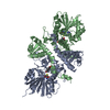
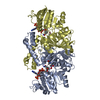
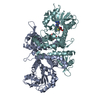
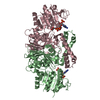
| Title | Crystal Structure Analysis of Caffeic acid/5-hydroxyferulic acid 3/5-O-methyltransferase Ferulic Acid Complex | ||||||
 Components Components | Caffeic acid 3-O-methyltransferase | ||||||
 Keywords Keywords | TRANSFERASE / O-methyltransferase / lignin / ferulic acid / methylation | ||||||
| Function / homology |  Function and homology information Function and homology informationcaffeate O-methyltransferase / caffeate O-methyltransferase activity / phenylpropanoid biosynthetic process / lignin biosynthetic process / S-adenosylmethionine-dependent methyltransferase activity / methylation / protein dimerization activity Similarity search - Function | ||||||
| Biological species |  | ||||||
| Method |  X-RAY DIFFRACTION / SYNCHROTRON / MOLECULAR REPLACEMENT / Resolution: 2.2 Å X-RAY DIFFRACTION / SYNCHROTRON / MOLECULAR REPLACEMENT / Resolution: 2.2 Å | ||||||
 Authors Authors | Zubieta, C. / Kota, P. / Ferrer, J.-L. / Dixon, R.A. / Noel, J.P. | ||||||
 Citation Citation | Journal: Plant Cell / Year: 2002 Title: Structural basis for the modulation of lignin monomer methylation by caffeic acid/5-hydroxyferulic acid 3/5-O-methyltransferase. Authors: Zubieta, C. / Kota, P. / Ferrer, J.L. / Dixon, R.A. / Noel, J.P. | ||||||
| History |
|
- Structure visualization
| Structure viewer | Molecule: MolmilJmol/JSmol |
|---|
- Downloads & links
-Download
| PDBx/mmCIF format | 1kyz.cif.gz | 219.5 KB | Display | PDBx/mmCIF format |
|---|---|---|---|---|
| PDB format | pdb1kyz.ent.gz | 176.3 KB | Display | PDB format |
| PDBx/mmJSON format | 1kyz.json.gz | Tree view | PDBx/mmJSON format | |
| Others |  Other downloads Other downloads |
-Validation report
| Arichive directory | https://data.pdbj.org/pub/pdb/validation_reports/ky/1kyzftp://data.pdbj.org/pub/pdb/validation_reports/ky/1kyz | HTTPS FTP |
|---|
-Related structure data


-Links
 PDBj
PDBj- Assembly
| Deposited unit | 
| ||||||||
|---|---|---|---|---|---|---|---|---|---|
| 1 | 
| ||||||||
| 2 | 
| ||||||||
| Unit cell |
| ||||||||
| Details | The biological assembly is a dimer of |
-Components
| #1: Protein | Mass: 39990.098 Da / Num. of mol.: 3 Source method: isolated from a genetically manipulated source Source: (gene. exp.)  #2: Chemical |   Mass: 194.184 Da / Num. of mol.: 3 / Source method: obtained synthetically / Formula: C10H10O4 Mass: 194.184 Da / Num. of mol.: 3 / Source method: obtained synthetically / Formula: C10H10O4#3: Chemical |   Type: L-peptide linking / Mass: 384.411 Da / Num. of mol.: 3 / Source method: obtained synthetically / Formula: C14H20N6O5S Type: L-peptide linking / Mass: 384.411 Da / Num. of mol.: 3 / Source method: obtained synthetically / Formula: C14H20N6O5S#4: Water | ChemComp-HOH / |  Mass: 18.015 Da / Num. of mol.: 264 / Source method: isolated from a natural source / Formula: H2O Mass: 18.015 Da / Num. of mol.: 264 / Source method: isolated from a natural source / Formula: H2O |
|---|
-Experimental details
-Experiment
| Experiment | Method: X-RAY DIFFRACTION / Number of used crystals: 1 |
|---|
- Sample preparation
| Crystal | Density Matthews: 2.75 Å3/Da / Density % sol: 55.33 % | |||||||||||||||||||||||||||||||||||||||||||||||||||||||||||||||
|---|---|---|---|---|---|---|---|---|---|---|---|---|---|---|---|---|---|---|---|---|---|---|---|---|---|---|---|---|---|---|---|---|---|---|---|---|---|---|---|---|---|---|---|---|---|---|---|---|---|---|---|---|---|---|---|---|---|---|---|---|---|---|---|---|
| Crystal grow | Temperature: 277 K / Method: vapor diffusion, hanging drop / pH: 8.5 Details: PEG 8000, calcium acetate, pH 8.5, VAPOR DIFFUSION, HANGING DROP, temperature 277K | |||||||||||||||||||||||||||||||||||||||||||||||||||||||||||||||
| Crystal grow | *PLUS Temperature: 4 ℃ / pH: 7.5 | |||||||||||||||||||||||||||||||||||||||||||||||||||||||||||||||
| Components of the solutions | *PLUS
|
-Data collection
| Diffraction | Mean temperature: 100 K |
|---|---|
| Diffraction source | Source: SYNCHROTRON / Site: SSRL  / Beamline: BL7-1 / Wavelength: 1.08 Å / Beamline: BL7-1 / Wavelength: 1.08 Å |
| Detector | Type: MARRESEARCH / Detector: IMAGE PLATE / Date: Feb 12, 2001 |
| Radiation | Monochromator: Si(111) / Protocol: SINGLE WAVELENGTH / Monochromatic (M) / Laue (L): M / Scattering type: x-ray |
| Radiation wavelength | Wavelength: 1.08 Å / Relative weight: 1 |
| Reflection | Resolution: 2.1→99 Å / Num. all: 68292 / Num. obs: 66087 / % possible obs: 96.8 % / Observed criterion σ(F): 2 / Observed criterion σ(I): 2 / Biso Wilson estimate: 16.4 Å2 |
| Reflection shell | Resolution: 2.19→2.23 Å / % possible all: 91 |
| Reflection | *PLUS Lowest resolution: 99 Å / Num. obs: 88232 / % possible obs: 98 % / Num. measured all: 388692 / Rmerge(I) obs: 0.049 |
| Reflection shell | *PLUS % possible obs: 99 % / Rmerge(I) obs: 0.51 |
- Processing
| Software |
| ||||||||||||||||||||||||||||||||||||
|---|---|---|---|---|---|---|---|---|---|---|---|---|---|---|---|---|---|---|---|---|---|---|---|---|---|---|---|---|---|---|---|---|---|---|---|---|---|
| Refinement | Method to determine structure: MOLECULAR REPLACEMENT Starting model: partial seleno-methionine model (unpublished) Resolution: 2.2→61.87 Å / Rfactor Rfree error: 0.005 / Data cutoff high absF: 1238884.41 / Data cutoff low absF: 0 / Isotropic thermal model: RESTRAINED / Cross valid method: THROUGHOUT / σ(F): 0 / Stereochemistry target values: Engh & Huber
| ||||||||||||||||||||||||||||||||||||
| Solvent computation | Solvent model: FLAT MODEL / Bsol: 48.1423 Å2 / ksol: 0.361389 e/Å3 | ||||||||||||||||||||||||||||||||||||
| Displacement parameters | Biso mean: 36.8 Å2
| ||||||||||||||||||||||||||||||||||||
| Refine analyze |
| ||||||||||||||||||||||||||||||||||||
| Refinement step | Cycle: LAST / Resolution: 2.2→61.87 Å
| ||||||||||||||||||||||||||||||||||||
| Refine LS restraints |
| ||||||||||||||||||||||||||||||||||||
| LS refinement shell | Resolution: 2.2→2.34 Å / Rfactor Rfree error: 0.013 / Total num. of bins used: 6
| ||||||||||||||||||||||||||||||||||||
| Xplor file |
| ||||||||||||||||||||||||||||||||||||
| Refinement | *PLUS Highest resolution: 2.1 Å / Lowest resolution: 99 Å / % reflection Rfree: 5 % / Rfactor obs: 0.236 / Rfactor Rfree: 0.274 / Rfactor Rwork: 0.236 | ||||||||||||||||||||||||||||||||||||
| Solvent computation | *PLUS | ||||||||||||||||||||||||||||||||||||
| Displacement parameters | *PLUS | ||||||||||||||||||||||||||||||||||||
| Refine LS restraints | *PLUS
| ||||||||||||||||||||||||||||||||||||
| LS refinement shell | *PLUS Rfactor Rfree: 0.315 / Rfactor Rwork: 0.267 |
