 Movie
Movie Controller

[English] 日本語
 Yorodumi
- PDB-7v1v: Difructose dianhydride I synthase/hydrolase (alphaFFase1) from Bi... -
+ Open data

- Basic information
| Entry | Database: PDB / ID: 7v1v | |||||||||
|---|---|---|---|---|---|---|---|---|---|---|
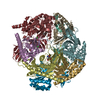
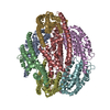
| Title | Difructose dianhydride I synthase/hydrolase (alphaFFase1) from Bifidobacterium dentium, ligand-free form | |||||||||
 Components Components | Difructose dianhydride I synthase/hydrolase (alphaFFase1) | |||||||||
 Keywords Keywords | HYDROLASE / Glycoside Hydrolase / alpha-D-fructofuranosidase / ligand-free | |||||||||
| Function / homology | GH172 domain-like / Protein of unknown function DUF2961 / GH172 second beta-sandwich domain / metal ion binding / D(-)-TARTARIC ACID / GH172 second beta-sandwich domain-containing protein Function and homology information Function and homology information | |||||||||
| Biological species |  Bifidobacterium dentium (bacteria) Bifidobacterium dentium (bacteria) | |||||||||
| Method |  X-RAY DIFFRACTION / SYNCHROTRON / MOLECULAR REPLACEMENT / Resolution: 1.96 Å X-RAY DIFFRACTION / SYNCHROTRON / MOLECULAR REPLACEMENT / Resolution: 1.96 Å | |||||||||
 Authors Authors | Kashima, T. / Arakawa, T. / Yamada, C. / Fujita, K. / Fushinobu, S. | |||||||||
| Funding support |  Japan, 2items Japan, 2items
| |||||||||
 Citation Citation | Journal: J.Biol.Chem. / Year: 2021 Title: Identification of difructose dianhydride I synthase/hydrolase from an oral bacterium establishes a novel glycoside hydrolase family. Authors: Kashima, T. / Okumura, K. / Ishiwata, A. / Kaieda, M. / Terada, T. / Arakawa, T. / Yamada, C. / Shimizu, K. / Tanaka, K. / Kitaoka, M. / Ito, Y. / Fujita, K. / Fushinobu, S. | |||||||||
| History |
|
- Structure visualization
| Structure viewer | Molecule: MolmilJmol/JSmol |
|---|
- Downloads & links
-Download
| PDBx/mmCIF format | 7v1v.cif.gz | 589.2 KB | Display | PDBx/mmCIF format |
|---|---|---|---|---|
| PDB format | pdb7v1v.ent.gz | 478.6 KB | Display | PDB format |
| PDBx/mmJSON format | 7v1v.json.gz | Tree view | PDBx/mmJSON format | |
| Others |  Other downloads Other downloads |
-Validation report
| Arichive directory | https://data.pdbj.org/pub/pdb/validation_reports/v1/7v1vftp://data.pdbj.org/pub/pdb/validation_reports/v1/7v1v | HTTPS FTP |
|---|
-Related structure data
| Related structure data | 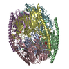 7v1wC 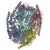 7v1xC 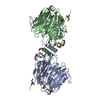 4kq7S S: Starting model for refinement C: citing same article ( |
|---|---|
| Similar structure data |


-Links
 PDBj
PDBj
- Assembly
| Deposited unit | 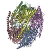
| ||||||||
|---|---|---|---|---|---|---|---|---|---|
| 1 |
| ||||||||
| Unit cell |
|
-Components
| #1: Protein | Mass: 53045.027 Da / Num. of mol.: 6 Source method: isolated from a genetically manipulated source Source: (gene. exp.) Bifidobacterium dentium (bacteria) / Gene: BDLFYP24_02130 / Plasmid: pET23d / Production host: #2: Chemical | ChemComp-CA /   Mass: 40.078 Da / Num. of mol.: 6 / Source method: obtained synthetically / Formula: Ca Mass: 40.078 Da / Num. of mol.: 6 / Source method: obtained synthetically / Formula: Ca#3: Chemical |   Mass: 118.174 Da / Num. of mol.: 2 / Source method: obtained synthetically / Formula: C6H14O2 / Comment: precipitant*YM Mass: 118.174 Da / Num. of mol.: 2 / Source method: obtained synthetically / Formula: C6H14O2 / Comment: precipitant*YM#4: Chemical |   Mass: 150.087 Da / Num. of mol.: 2 / Source method: obtained synthetically / Formula: C4H6O6 Mass: 150.087 Da / Num. of mol.: 2 / Source method: obtained synthetically / Formula: C4H6O6#5: Water | ChemComp-HOH / |  Mass: 18.015 Da / Num. of mol.: 2423 / Source method: isolated from a natural source / Formula: H2O Mass: 18.015 Da / Num. of mol.: 2423 / Source method: isolated from a natural source / Formula: H2OHas ligand of interest | N | |
|---|
-Experimental details
-Experiment
| Experiment | Method: X-RAY DIFFRACTION / Number of used crystals: 1 |
|---|
- Sample preparation
| Crystal | Density Matthews: 2.39 Å3/Da / Density % sol: 48.45 % |
|---|---|
| Crystal grow | Temperature: 293.15 K / Method: vapor diffusion, sitting drop / Details: 20% PEG3350, 0.2M di-sodium tartrate |
-Data collection
| Diffraction | Mean temperature: 100 K / Serial crystal experiment: N |
|---|---|
| Diffraction source | Source: SYNCHROTRON / Site: Photon Factory / Beamline: BL-5A / Wavelength: 1 Å |
| Detector | Type: DECTRIS PILATUS 6M / Detector: PIXEL / Date: Jun 24, 2019 |
| Radiation | Monochromator: Numerical link type Si(111) double crystal / Protocol: SINGLE WAVELENGTH / Monochromatic (M) / Laue (L): M / Scattering type: x-ray |
| Radiation wavelength | Wavelength: 1 Å / Relative weight: 1 |
| Reflection | Resolution: 1.96→49.69 Å / Num. obs: 206183 / % possible obs: 99.9 % / Redundancy: 3.5 % / Biso Wilson estimate: 8.99 Å2 / CC1/2: 0.994 / Rmerge(I) obs: 0.098 / Rpim(I) all: 0.061 / Rrim(I) all: 0.116 / Net I/σ(I): 10.3 |
| Reflection shell | Resolution: 1.96→1.99 Å / Redundancy: 3.2 % / Rmerge(I) obs: 0.432 / Mean I/σ(I) obs: 2.9 / Num. unique obs: 10181 / CC1/2: 0.8 / Rpim(I) all: 0.283 / Rrim(I) all: 0.518 / % possible all: 99.9 |
- Processing
| Software |
| ||||||||||||||||||||||||||||||||||||||||||||||||||||||||||||
|---|---|---|---|---|---|---|---|---|---|---|---|---|---|---|---|---|---|---|---|---|---|---|---|---|---|---|---|---|---|---|---|---|---|---|---|---|---|---|---|---|---|---|---|---|---|---|---|---|---|---|---|---|---|---|---|---|---|---|---|---|---|
| Refinement | Method to determine structure: MOLECULAR REPLACEMENT Starting model: 4KQ7 Resolution: 1.96→49.69 Å / Cor.coef. Fo:Fc: 0.96 / Cor.coef. Fo:Fc free: 0.94 / SU B: 3.421 / SU ML: 0.096 / Cross valid method: THROUGHOUT / σ(F): 0 / ESU R: 0.151 / ESU R Free: 0.134 / Stereochemistry target values: MAXIMUM LIKELIHOOD Details: HYDROGENS HAVE BEEN ADDED IN THE RIDING POSITIONS U VALUES : REFINED INDIVIDUALLY
| ||||||||||||||||||||||||||||||||||||||||||||||||||||||||||||
| Solvent computation | Ion probe radii: 0.8 Å / Shrinkage radii: 0.8 Å / VDW probe radii: 1.2 Å / Solvent model: MASK | ||||||||||||||||||||||||||||||||||||||||||||||||||||||||||||
| Displacement parameters | Biso max: 91.27 Å2 / Biso mean: 18.101 Å2 / Biso min: 5.38 Å2
| ||||||||||||||||||||||||||||||||||||||||||||||||||||||||||||
| Refinement step | Cycle: final / Resolution: 1.96→49.69 Å
| ||||||||||||||||||||||||||||||||||||||||||||||||||||||||||||
| Refine LS restraints |
| ||||||||||||||||||||||||||||||||||||||||||||||||||||||||||||
| LS refinement shell | Resolution: 1.96→2.01 Å / Rfactor Rfree error: 0
|
