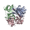
- PDB-4kq7: Crystal structure of a DUF2961 family protein (BACUNI_00161) from... -
+
Open data
ID or keywords:
Loading...
-
Basic information
Entry
Database: PDB / ID: 4kq7
Title
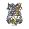
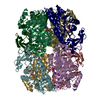
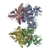
Crystal structure of a DUF2961 family protein (BACUNI_00161) from Bacteroides uniformis ATCC 8492 at 1.62 A resolution
Components
hypothetical protein
Keywords
Structural Genomics / Unknown Function / PF11175 family protein / DUF2961 / Joint Center for Structural Genomics / JCSG / Protein Structure Initiative / PSI-BIOLOGY
Function / homology
Protein of unknown function DUF2961 / Protein of unknown function DUF2961 / GH172 second beta-sandwich domain / Jelly Rolls / Sandwich / Mainly Beta / DI(HYDROXYETHYL)ETHER / DUF2961 domain-containing protein
Function and homology information
Biological species
Bacteroides uniformis (bacteria)
Method
X-RAY DIFFRACTION / SYNCHROTRON / SAD / Resolution: 1.62 Å
Mass: 18.015 Da / Num. of mol.: 878 / Source method: isolated from a natural source / Formula: H2O
-
Details
Has protein modification
Y
Sequence details
THE CONSTRUCT WAS EXPRESSED WITH A PURIFICATION TAG MGSDKIHHHHHHENLYFQG. THE TAG WAS REMOVED WITH ...THE CONSTRUCT WAS EXPRESSED WITH A PURIFICATION TAG MGSDKIHHHHHHENLYFQG. THE TAG WAS REMOVED WITH TEV PROTEASE LEAVING ONLY A GLYCINE (0) FOLLOWED BY RESIDUES 45-417 OF THE TARGET SEQUENCE.
-
Experimental details
-
Experiment
Experiment
Method: X-RAY DIFFRACTION / Number of used crystals: 1
-
Sample preparation
Crystal
Density Matthews: 2.32 Å3/Da / Density % sol: 46.93 %
Type: DECTRIS PILATUS 6M / Detector: PIXEL / Date: Jan 24, 2013 Details: Flat mirror (vertical focusing); single crystal Si(111) bent monochromator (horizontal focusing)
Radiation
Monochromator: single crystal Si(111) bent / Protocol: SAD / Monochromatic (M) / Laue (L): M / Scattering type: x-ray
Radiation wavelength
Wavelength: 0.97858 Å / Relative weight: 1
Reflection
Resolution: 1.62→47.35 Å / Num. obs: 101280 / % possible obs: 99.9 % / Observed criterion σ(I): -3 / Biso Wilson estimate: 17.374 Å2 / Rmerge(I) obs: 0.098 / Net I/σ(I): 12.73
Reflection shell
Diffraction-ID: 1
Resolution (Å)
Highest resolution (Å)
Rmerge(I) obs
Mean I/σ(I) obs
Num. measured obs
Num. unique obs
% possible all
1.62-1.68
0.821
2.3
72900
10352
100
1.68-1.75
0.642
2.9
72141
10422
99.9
1.75-1.82
0.453
4
57190
8811
99.9
1.82-1.92
0.318
5.8
74853
10556
100
1.92-2.04
0.214
8.3
70788
10062
100
2.04-2.2
0.151
11.2
67638
10243
99.8
2.2-2.42
0.117
14.6
71803
10030
100
2.42-2.77
0.094
17.8
70242
10149
100
2.77-3.48
0.061
25.8
70775
10099
100
3.48
0.046
33.3
70014
10547
99.8
-
Phasing
Phasing
Method: SAD
-
Processing
Software
Name
Version
Classification
NB
MolProbity
3beta29
modelbuilding
PDB_EXTRACT
3.1
dataextraction
SHELX
phasing
SHARP
phasing
XSCALE
July4, 2012
datascaling
BUSTER-TNT
2.10.0
refinement
XDS
datareduction
SHELXD
phasing
BUSTER
2.10.0
refinement
Refinement
Method to determine structure: SAD / Resolution: 1.62→47.35 Å / Cor.coef. Fo:Fc: 0.9728 / Cor.coef. Fo:Fc free: 0.9685 / Occupancy max: 1 / Occupancy min: 0.15 / Cross valid method: THROUGHOUT / σ(F): 0 Details: 1. ATOM RECORD CONTAINS SUM OF TLS AND RESIDUAL B FACTORS. ANISOU RECORD CONTAINS SUM OF TLS AND RESIDUAL U FACTORS. 2. A MET-INHIBITION PROTOCOL WAS USED FOR SELENOMETHIONINE INCORPORATION ...Details: 1. ATOM RECORD CONTAINS SUM OF TLS AND RESIDUAL B FACTORS. ANISOU RECORD CONTAINS SUM OF TLS AND RESIDUAL U FACTORS. 2. A MET-INHIBITION PROTOCOL WAS USED FOR SELENOMETHIONINE INCORPORATION DURING PROTEIN EXPRESSION. THE OCCUPANCY OF THE SE ATOMS IN THE MSE RESIDUES WAS REDUCED TO 0.75 TO ACCOUNT FOR THE REDUCED SCATTERING POWER DUE TO PARTIAL S-MET INCORPORATION. 3. POLYETHYLENE GLYCOL AND CL-ION FROM THE CRYOPROTECTANT AND FROM THE CRYSTALLIZATION CONDITION HAVE BEEN MODELED IN THE SOLVENT STRUCTURE. 4. THE MAD PHASES WERE USED AS RESTRAINTS DURING REFINEMENT. 5.NCS RESTRAINTS WERE IMPOSED BY AUTOBUSTER'S LSSR PROCEDURE (-AUTONCS).
In the structure databanks used in Yorodumi, some data are registered as the other names, "COVID-19 virus" and "2019-nCoV". Here are the details of the virus and the list of structure data.
Jan 31, 2019. EMDB accession codes are about to change! (news from PDBe EMDB page)
EMDB accession codes are about to change! (news from PDBe EMDB page)
The allocation of 4 digits for EMDB accession codes will soon come to an end. Whilst these codes will remain in use, new EMDB accession codes will include an additional digit and will expand incrementally as the available range of codes is exhausted. The current 4-digit format prefixed with “EMD-” (i.e. EMD-XXXX) will advance to a 5-digit format (i.e. EMD-XXXXX), and so on. It is currently estimated that the 4-digit codes will be depleted around Spring 2019, at which point the 5-digit format will come into force.
The EM Navigator/Yorodumi systems omit the EMD- prefix.
Related info.:Q: What is EMD? / ID/Accession-code notation in Yorodumi/EM Navigator
Yorodumi is a browser for structure data from EMDB, PDB, SASBDB, etc.
This page is also the successor to EM Navigator detail page, and also detail information page/front-end page for Omokage search.
The word "yorodu" (or yorozu) is an old Japanese word meaning "ten thousand". "mi" (miru) is to see.
Related info.:EMDB / PDB / SASBDB / Comparison of 3 databanks / Yorodumi Search / Aug 31, 2016. New EM Navigator & Yorodumi / Yorodumi Papers / Jmol/JSmol / Function and homology information / Changes in new EM Navigator and Yorodumi
 Movie
Movie Controller

 Yorodumi
 Open data
 Basic information
 Components
 Keywords
 Function and homology information
 Bacteroides uniformis (bacteria)
 X-RAY DIFFRACTION /
  Authors
 Citation
 Structure visualization
 Downloads & links
 Other downloads
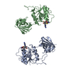

 PDBj
PDBj Assembly
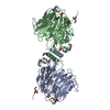


 Mass: 22.990 Da / Num. of mol.: 7 / Source method: obtained synthetically / Formula: Na
 Mass: 194.226 Da / Num. of mol.: 4 / Source method: obtained synthetically / Formula: C8H18O5 / Comment: precipitant*YM
 Mass: 106.120 Da / Num. of mol.: 3 / Source method: obtained synthetically / Formula: C4H10O3
 Mass: 238.278 Da / Num. of mol.: 1 / Source method: obtained synthetically / Formula: C10H22O6 / Comment: precipitant*YM
 Sample preparation
 / Beamline: BL11-1 / Wavelength: 0.97858
  Processing