 Movie
Movie Controller


+ Open data

- Basic information
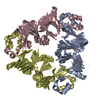
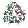
| Entry | Database: PDB / ID: 7u1h | ||||||
|---|---|---|---|---|---|---|---|
| Title | Crystal structure of Lens culinaris vicilin | ||||||
 Components Components | Allergen Len c 1.0101 | ||||||
 Keywords Keywords | ALLERGEN / Cupin / Vicilin / Convicilin / Lentil / Seed Storage Protein | ||||||
| Function / homology |  Function and homology information Function and homology information | ||||||
| Biological species |  Lens culinaris (lentil) Lens culinaris (lentil) | ||||||
| Method |  X-RAY DIFFRACTION / SYNCHROTRON / MOLECULAR REPLACEMENT / Resolution: 2.5 Å X-RAY DIFFRACTION / SYNCHROTRON / MOLECULAR REPLACEMENT / Resolution: 2.5 Å | ||||||
 Authors Authors | Robinson, K.A. / Bakestani, I.D. / Loewen, M.C. | ||||||
| Funding support |  Canada, 1items Canada, 1items
| ||||||
 Citation Citation | Journal: Food Chem (Oxf) / Year: 2022 Title: Pea and lentil 7S globulin crystal structures with comparative immunoglobulin epitope mapping. Authors: Robinson, K.A. / St-Jacques, A.D. / Bakestani, I.D. / Beavington, B.A.G. / Loewen, M.C. | ||||||
| History |
|
- Structure visualization
| Structure viewer | Molecule: MolmilJmol/JSmol |
|---|
- Downloads & links
-Download
| PDBx/mmCIF format | 7u1h.cif.gz | 433.6 KB | Display | PDBx/mmCIF format |
|---|---|---|---|---|
| PDB format | pdb7u1h.ent.gz | 345.5 KB | Display | PDB format |
| PDBx/mmJSON format | 7u1h.json.gz | Tree view | PDBx/mmJSON format | |
| Others |  Other downloads Other downloads |
-Validation report
| Arichive directory | https://data.pdbj.org/pub/pdb/validation_reports/u1/7u1hftp://data.pdbj.org/pub/pdb/validation_reports/u1/7u1h | HTTPS FTP |
|---|
-Related structure data
| Related structure data | 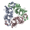 7u1iC  7u1jC  1uikS S: Starting model for refinement C: citing same article ( |
|---|---|
| Similar structure data |


-Links
 PDBj
PDBj- Assembly
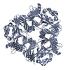
| Deposited unit | 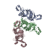
| ||||||||
|---|---|---|---|---|---|---|---|---|---|
| 1 | 
| ||||||||
| Unit cell |
|
-Components
| #1: Protein | Mass: 49377.871 Da / Num. of mol.: 3 Source method: isolated from a genetically manipulated source Source: (gene. exp.) Lens culinaris (lentil)Production host:  References: UniProt: Q84UI1 #2: Water | ChemComp-HOH / |  Mass: 18.015 Da / Num. of mol.: 40 / Source method: isolated from a natural source / Formula: H2O Mass: 18.015 Da / Num. of mol.: 40 / Source method: isolated from a natural source / Formula: H2O |
|---|
-Experimental details
-Experiment
| Experiment | Method: X-RAY DIFFRACTION / Number of used crystals: 1 |
|---|
- Sample preparation
| Crystal | Density Matthews: 2.02 Å3/Da / Density % sol: 39.25 % |
|---|---|
| Crystal grow | Temperature: 293 K / Method: vapor diffusion, hanging drop / pH: 5.5 Details: 0.01 M Zinc Chloride, 0.1 M Sodium acetate trihydrate, 15% PEG6000, with 20% glycerol as cryoprotectant |
-Data collection
| Diffraction | Mean temperature: 100 K / Serial crystal experiment: N |
|---|---|
| Diffraction source | Source: SYNCHROTRON / Site: CLSI / Beamline: 08B1-1 / Wavelength: 0.98 Å |
| Detector | Type: DECTRIS PILATUS3 S 6M / Detector: PIXEL / Date: Aug 18, 2021 |
| Radiation | Protocol: SINGLE WAVELENGTH / Monochromatic (M) / Laue (L): M / Scattering type: x-ray |
| Radiation wavelength | Wavelength: 0.98 Å / Relative weight: 1 |
| Reflection | Resolution: 2.5→143.74 Å / Num. obs: 39945 / % possible obs: 98.6 % / Redundancy: 6.2 % / CC1/2: 0.991 / Rmerge(I) obs: 0.144 / Rpim(I) all: 0.093 / Rrim(I) all: 0.172 / Net I/σ(I): 9.6 |
| Reflection shell | Resolution: 2.5→2.6 Å / Redundancy: 6.2 % / Rmerge(I) obs: 0.782 / Num. unique obs: 4426 / CC1/2: 0.812 / Rpim(I) all: 0.513 / Rrim(I) all: 0.939 |
- Processing
| Software |
| |||||||||||||||||||||||||||||||||||||||||||||||||||||||||||||||||||||||||||||||||||||||||||||||||||||||||||||||||||||||||||||||||||||||||||||||||||||||||||
|---|---|---|---|---|---|---|---|---|---|---|---|---|---|---|---|---|---|---|---|---|---|---|---|---|---|---|---|---|---|---|---|---|---|---|---|---|---|---|---|---|---|---|---|---|---|---|---|---|---|---|---|---|---|---|---|---|---|---|---|---|---|---|---|---|---|---|---|---|---|---|---|---|---|---|---|---|---|---|---|---|---|---|---|---|---|---|---|---|---|---|---|---|---|---|---|---|---|---|---|---|---|---|---|---|---|---|---|---|---|---|---|---|---|---|---|---|---|---|---|---|---|---|---|---|---|---|---|---|---|---|---|---|---|---|---|---|---|---|---|---|---|---|---|---|---|---|---|---|---|---|---|---|---|---|---|---|
| Refinement | Method to determine structure: MOLECULAR REPLACEMENT Starting model: 1UIK Resolution: 2.5→77.909 Å / Cor.coef. Fo:Fc: 0.939 / Cor.coef. Fo:Fc free: 0.878 / WRfactor Rfree: 0.254 / WRfactor Rwork: 0.181 / SU B: 12.975 / SU ML: 0.281 / Average fsc free: 0.8585 / Average fsc work: 0.8906 / Cross valid method: NONE / ESU R: 0.939 / ESU R Free: 0.351 Details: Hydrogens have been added in their riding positions
| |||||||||||||||||||||||||||||||||||||||||||||||||||||||||||||||||||||||||||||||||||||||||||||||||||||||||||||||||||||||||||||||||||||||||||||||||||||||||||
| Solvent computation | Ion probe radii: 0.8 Å / Shrinkage radii: 0.8 Å / VDW probe radii: 1.2 Å / Solvent model: MASK BULK SOLVENT | |||||||||||||||||||||||||||||||||||||||||||||||||||||||||||||||||||||||||||||||||||||||||||||||||||||||||||||||||||||||||||||||||||||||||||||||||||||||||||
| Displacement parameters | Biso mean: 36.052 Å2
| |||||||||||||||||||||||||||||||||||||||||||||||||||||||||||||||||||||||||||||||||||||||||||||||||||||||||||||||||||||||||||||||||||||||||||||||||||||||||||
| Refinement step | Cycle: LAST / Resolution: 2.5→77.909 Å
| |||||||||||||||||||||||||||||||||||||||||||||||||||||||||||||||||||||||||||||||||||||||||||||||||||||||||||||||||||||||||||||||||||||||||||||||||||||||||||
| Refine LS restraints |
| |||||||||||||||||||||||||||||||||||||||||||||||||||||||||||||||||||||||||||||||||||||||||||||||||||||||||||||||||||||||||||||||||||||||||||||||||||||||||||
| LS refinement shell |
|
