 Movie
Movie Controller

[English] 日本語
 Yorodumi
- PDB-7thz: Structure of Leucine Rich Repeat Kinase 2's ROC domain interactin... -
+ Open data

- Basic information
| Entry | Database: PDB / ID: 7thz | |||||||||||||||
|---|---|---|---|---|---|---|---|---|---|---|---|---|---|---|---|---|
| Title | Structure of Leucine Rich Repeat Kinase 2's ROC domain interacting with the microtubule facing the plus end | |||||||||||||||
 Components Components | Leucine-rich repeat serine/threonine-protein kinase 2 | |||||||||||||||
 Keywords Keywords | CYTOSOLIC PROTEIN / parkinson's disease / microtubule / kinase / gtpase | |||||||||||||||
| Function / homology |  Function and homology information Function and homology informationcaveola neck / : / beta-catenin destruction complex binding / regulation of branching morphogenesis of a nerve / Wnt signalosome assembly / negative regulation of motile cilium assembly / regulation of neuron maturation / regulation of kidney size / regulation of cell projection organization / regulation of dopamine receptor signaling pathway ...caveola neck / : / beta-catenin destruction complex binding / regulation of branching morphogenesis of a nerve / Wnt signalosome assembly / negative regulation of motile cilium assembly / regulation of neuron maturation / regulation of kidney size / regulation of cell projection organization / regulation of dopamine receptor signaling pathway / tangential migration from the subventricular zone to the olfactory bulb / GTP-dependent protein kinase activity / regulation of SNARE complex assembly / regulation of neuroblast proliferation / regulation of ER to Golgi vesicle-mediated transport / protein localization to endoplasmic reticulum exit site / negative regulation of late endosome to lysosome transport / regulation of mitochondrial depolarization / : / peroxidase inhibitor activity / positive regulation of dopamine receptor signaling pathway / amphisome / regulation of synaptic vesicle transport / : / regulation of CAMKK-AMPK signaling cascade / negative regulation of autophagosome assembly / co-receptor binding / positive regulation of microglial cell activation / olfactory bulb development / regulation of retrograde transport, endosome to Golgi / negative regulation of GTPase activity / cellular response to curcumin / striatum development / regulation of locomotion / positive regulation of synaptic vesicle endocytosis / cytoplasmic side of mitochondrial outer membrane / JUN kinase kinase kinase activity / regulation of cAMP/PKA signal transduction / multivesicular body, internal vesicle / negative regulation of excitatory postsynaptic potential / endoplasmic reticulum organization / neuron projection arborization / mitochondrion localization / regulation of dendritic spine morphogenesis / protein localization to mitochondrion / positive regulation of mitochondrial outer membrane permeabilization involved in apoptotic signaling pathway / cellular response to dopamine / negative regulation of protein processing / positive regulation of protein autoubiquitination / Wnt signalosome / exploration behavior / GTP metabolic process / regulation of reactive oxygen species metabolic process / regulation of canonical Wnt signaling pathway / lysosome organization / syntaxin-1 binding / positive regulation of programmed cell death / negative regulation of macroautophagy / Golgi-associated vesicle / PTK6 promotes HIF1A stabilization / clathrin binding / Golgi organization / regulation of synaptic vesicle exocytosis / regulation of mitochondrial fission / Lewy body / intracellular distribution of mitochondria / protein kinase A binding / microvillus / canonical Wnt signaling pathway / neuromuscular junction development / autolysosome / locomotory exploration behavior / endoplasmic reticulum exit site / MAP kinase kinase kinase activity / JNK cascade / regulation of synaptic vesicle endocytosis / negative regulation of endoplasmic reticulum stress-induced intrinsic apoptotic signaling pathway / Rho protein signal transduction / regulation of synaptic transmission, glutamatergic / presynaptic cytosol / determination of adult lifespan / cellular response to manganese ion / neuron projection morphogenesis / phagocytic vesicle / positive regulation of autophagy / dendrite cytoplasm / excitatory postsynaptic potential / positive regulation of protein ubiquitination / cellular response to starvation / regulation of autophagy / GTPase activator activity / mitochondrion organization / calcium-mediated signaling / SNARE binding / cellular response to reactive oxygen species / trans-Golgi network / regulation of protein stability / regulation of membrane potential / tubulin binding / mitochondrial membrane Similarity search - Function | |||||||||||||||
| Biological species |  Homo sapiens (human) Homo sapiens (human) | |||||||||||||||
| Method | ELECTRON MICROSCOPY / single particle reconstruction / cryo EM / Resolution: 5 Å | |||||||||||||||
 Authors Authors | Matyszewski, M. / Leschziner, A.E. | |||||||||||||||
| Funding support |  United States, 4items United States, 4items
| |||||||||||||||
 Citation Citation | Journal: Nat Struct Mol Biol / Year: 2022 Title: Structural basis for Parkinson's disease-linked LRRK2's binding to microtubules. Authors: David M Snead / Mariusz Matyszewski / Andrea M Dickey / Yu Xuan Lin / Andres E Leschziner / Samara L Reck-Peterson / Abstract: Leucine-rich repeat kinase 2 (LRRK2) is one of the most commonly mutated genes in familial Parkinson's disease (PD). Under some circumstances, LRRK2 co-localizes with microtubules in cells, an ...Leucine-rich repeat kinase 2 (LRRK2) is one of the most commonly mutated genes in familial Parkinson's disease (PD). Under some circumstances, LRRK2 co-localizes with microtubules in cells, an association enhanced by PD mutations. We report a cryo-EM structure of the catalytic half of LRRK2, containing its kinase, in a closed conformation, and GTPase domains, bound to microtubules. We also report a structure of the catalytic half of LRRK1, which is closely related to LRRK2 but is not linked to PD. Although LRRK1's structure is similar to that of LRRK2, we find that LRRK1 does not interact with microtubules. Guided by these structures, we identify amino acids in LRRK2's GTPase that mediate microtubule binding; mutating them disrupts microtubule binding in vitro and in cells, without affecting LRRK2's kinase activity. Our results have implications for the design of therapeutic LRRK2 kinase inhibitors. | |||||||||||||||
| History |
|
- Structure visualization
| Structure viewer | Molecule: MolmilJmol/JSmol |
|---|
- Downloads & links
-Download
| PDBx/mmCIF format | 7thz.cif.gz | 148.7 KB | Display | PDBx/mmCIF format |
|---|---|---|---|---|
| PDB format | pdb7thz.ent.gz | 107.2 KB | Display | PDB format |
| PDBx/mmJSON format | 7thz.json.gz | Tree view | PDBx/mmJSON format | |
| Others |  Other downloads Other downloads |
-Validation report
| Arichive directory | https://data.pdbj.org/pub/pdb/validation_reports/th/7thzftp://data.pdbj.org/pub/pdb/validation_reports/th/7thz | HTTPS FTP |
|---|
-Related structure data
| Related structure data |  25907MC 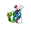 7thyC C: citing same article ( M: map data used to model this data |
|---|---|
| Similar structure data |


-Links
 PDBj
PDBj


- Assembly
| Deposited unit | 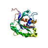
|
|---|---|
| 1 |
|
| Number of models | 5 |
-Components
| #1: Protein | Mass: 22191.762 Da / Num. of mol.: 1 / Fragment: ROC domain Source method: isolated from a genetically manipulated source Source: (gene. exp.) Homo sapiens (human) / Gene: LRRK2, PARK8 / Production host:   Spodoptera frugiperda (fall armyworm) Spodoptera frugiperda (fall armyworm)References: UniProt: Q5S007, non-specific serine/threonine protein kinase, Hydrolases; Acting on acid anhydrides; Acting on GTP to facilitate cellular and subcellular movement |
|---|---|
| #2: Chemical | ChemComp-GDP /   Type: RNA linking / Mass: 443.201 Da / Num. of mol.: 1 / Source method: obtained synthetically / Formula: C10H15N5O11P2 / Comment: GDP, energy-carrying molecule*YM Type: RNA linking / Mass: 443.201 Da / Num. of mol.: 1 / Source method: obtained synthetically / Formula: C10H15N5O11P2 / Comment: GDP, energy-carrying molecule*YM |
| Has ligand of interest | N |
-Experimental details
-Experiment
| Experiment | Method: ELECTRON MICROSCOPY |
|---|---|
| EM experiment | Aggregation state: FILAMENT / 3D reconstruction method: single particle reconstruction |
- Sample preparation
| Component | Name: LRRK2RCKW filament bound to a 11-pf microtubule with MLi-2 present Type: COMPLEX / Entity ID: #1 / Source: MULTIPLE SOURCES | ||||||||||||||||||||||||||||||
|---|---|---|---|---|---|---|---|---|---|---|---|---|---|---|---|---|---|---|---|---|---|---|---|---|---|---|---|---|---|---|---|
| Molecular weight | Experimental value: NO | ||||||||||||||||||||||||||||||
| Buffer solution | pH: 7.4 Details: This is the final dilution buffer. The incubation buffer consisted of 1x BRB80, 10% glycerol, 1mM DTT, 1mM GTP, 1mM MgCl2, 10 uM taxol, and 5 uM MLi-2. Sample was diluted 3-fold right before ...Details: This is the final dilution buffer. The incubation buffer consisted of 1x BRB80, 10% glycerol, 1mM DTT, 1mM GTP, 1mM MgCl2, 10 uM taxol, and 5 uM MLi-2. Sample was diluted 3-fold right before freezing with the final buffer. | ||||||||||||||||||||||||||||||
| Buffer component |
| ||||||||||||||||||||||||||||||
| Specimen | Embedding applied: NO / Shadowing applied: NO / Staining applied: NO / Vitrification applied: YES Details: 4.5 uM of LRRK2RCKW (I2020T) was allowed to incubate with 2.25 uM of tubulin dimer, causing both to co-polymerize. 5 uM of MLi-2 was present as well. The sample was diluted 3-fold right ...Details: 4.5 uM of LRRK2RCKW (I2020T) was allowed to incubate with 2.25 uM of tubulin dimer, causing both to co-polymerize. 5 uM of MLi-2 was present as well. The sample was diluted 3-fold right before freezing (1.5 uM LRRK2RCKW concentration final). | ||||||||||||||||||||||||||||||
| Specimen support | Details: EMS LC-300 lacey grid used (not homemade, but can't choose EMS as the manufacturer) Grid material: COPPER / Grid mesh size: 300 divisions/in. / Grid type: Homemade | ||||||||||||||||||||||||||||||
| Vitrification | Cryogen name: ETHANE / Humidity: 100 % / Chamber temperature: 277 K |
- Electron microscopy imaging
| Experimental equipment |  Model: Talos Arctica / Image courtesy: FEI Company |
|---|---|
| Microscopy | Model: FEI TALOS ARCTICA |
| Electron gun | Electron source:  FIELD EMISSION GUN / Accelerating voltage: 200 kV / Illumination mode: FLOOD BEAM FIELD EMISSION GUN / Accelerating voltage: 200 kV / Illumination mode: FLOOD BEAM |
| Electron lens | Mode: BRIGHT FIELD / Nominal magnification: 36000 X / Nominal defocus max: 1500 nm / Nominal defocus min: 1500 nm / Cs: 2.7 mm / C2 aperture diameter: 70 µm / Alignment procedure: COMA FREE |
| Specimen holder | Cryogen: NITROGEN |
| Image recording | Average exposure time: 10 sec. / Electron dose: 55 e/Å2 / Detector mode: COUNTING / Film or detector model: GATAN K2 SUMMIT (4k x 4k) / Num. of grids imaged: 2 / Details: 250 ms frames |
- Processing
| EM software |
| ||||||||||||||||||||||||||||||||||||||||
|---|---|---|---|---|---|---|---|---|---|---|---|---|---|---|---|---|---|---|---|---|---|---|---|---|---|---|---|---|---|---|---|---|---|---|---|---|---|---|---|---|---|
| CTF correction | Type: PHASE FLIPPING AND AMPLITUDE CORRECTION | ||||||||||||||||||||||||||||||||||||||||
| Particle selection | Num. of particles selected: 557577 Details: Filament Autopicker with templates created by manual picking. This is before symmetry expansion. | ||||||||||||||||||||||||||||||||||||||||
| Symmetry | Point symmetry: C1 (asymmetric) | ||||||||||||||||||||||||||||||||||||||||
| 3D reconstruction | Resolution: 5 Å / Resolution method: FSC 0.143 CUT-OFF / Num. of particles: 99854 / Symmetry type: POINT | ||||||||||||||||||||||||||||||||||||||||
| Atomic model building | Protocol: FLEXIBLE FIT Details: Used AlphaFold model as initial model (Q5S007) using only the ROC domain. TUB1 was added to the initial refinement to prevent ROC model from entering density reserved for the microtubule. ...Details: Used AlphaFold model as initial model (Q5S007) using only the ROC domain. TUB1 was added to the initial refinement to prevent ROC model from entering density reserved for the microtubule. TUB1 was discarded after the initial refinement. |
