 Movie
Movie Controller

[English] 日本語
 Yorodumi
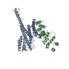
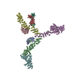
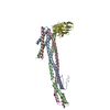
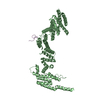
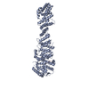
- PDB-7tbl: Composite structure of the human nuclear pore complex (NPC) cytop... -
+ Open data

- Basic information
| Entry | Database: PDB / ID: 7tbl | |||||||||||||||
|---|---|---|---|---|---|---|---|---|---|---|---|---|---|---|---|---|
| Title | Composite structure of the human nuclear pore complex (NPC) cytoplasmic face generated with a 12A cryo-ET map of the purified HeLa cell NPC | |||||||||||||||
 Components Components |
| |||||||||||||||
 Keywords Keywords | TRANSPORT PROTEIN / nuclear pore complex / nucleocytoplasmic transport / alpha-helical solenoid / nuclear pore | |||||||||||||||
| Biological species |  Homo sapiens (human) Homo sapiens (human) | |||||||||||||||
| Method | ELECTRON MICROSCOPY / subtomogram averaging / cryo EM / Resolution: 23 Å | |||||||||||||||
 Authors Authors | Bley, C.J. / Nie, S. / Mobbs, G.W. / Petrovic, S. / Gres, A.T. / Liu, X. / Mukherjee, S. / Harvey, S. / Huber, F.M. / Lin, D.H. ...Bley, C.J. / Nie, S. / Mobbs, G.W. / Petrovic, S. / Gres, A.T. / Liu, X. / Mukherjee, S. / Harvey, S. / Huber, F.M. / Lin, D.H. / Brown, B. / Tang, A.W. / Rundlet, E.J. / Correia, A.R. / Chen, S. / Regmi, S.G. / Stevens, T.A. / Jette, C.A. / Dasso, M. / Patke, A. / Palazzo, A.F. / Kossiakoff, A.A. / Hoelz, A. | |||||||||||||||
| Funding support |  United States, 4items United States, 4items
| |||||||||||||||
 Citation Citation | Journal: Science / Year: 2022 Title: AI-based structure prediction empowers integrative structural analysis of human nuclear pores Authors: Mosalaganti, S. / Obarska-Kosinska, A. / Siggel, M. / Taniguchi, R. / Turonova, B. / Zimmerli, C.E. / Buczak, K. / Schmidt, F.H. / Margiotta, E. / Mackmull, M.-T. / Hagen, W.J.H. / Hummer, G. ...Authors: Mosalaganti, S. / Obarska-Kosinska, A. / Siggel, M. / Taniguchi, R. / Turonova, B. / Zimmerli, C.E. / Buczak, K. / Schmidt, F.H. / Margiotta, E. / Mackmull, M.-T. / Hagen, W.J.H. / Hummer, G. / Kosinski, J. / Beck, M. | |||||||||||||||
| History |
| |||||||||||||||
| Remark 0 | THIS ENTRY 7TBL REFLECTS AN ALTERNATIVE MODELING OF THE ORIGINAL DATA IN EMD-14322, DETERMINED BY S. ...THIS ENTRY 7TBL REFLECTS AN ALTERNATIVE MODELING OF THE ORIGINAL DATA IN EMD-14322, DETERMINED BY S.Mosalaganti,A.Obarska-Kosinska,M.Siggel,R.Taniguchi,B.Turonova, C.E.Zimmerli,K.Buczak,F.H.Schmidt,E.Margiotta,M.T.Mackmull,W.J.H.Hagen,G.Hummer,J.Kosinski,M.Beck |
- Structure visualization
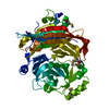
| Structure viewer | Molecule:  MolmilJmol/JSmol MolmilJmol/JSmol |
|---|
- Downloads & links
-Download
| PDBx/mmCIF format | 7tbl.cif.gz | 9.6 MB | Display | PDBx/mmCIF format |
|---|---|---|---|---|
| PDB format | pdb7tbl.ent.gz | Display | PDB format | |
| PDBx/mmJSON format | 7tbl.json.gz | Tree view | PDBx/mmJSON format | |
| Others |  Other downloads Other downloads |
-Validation report
| Arichive directory | https://data.pdbj.org/pub/pdb/validation_reports/tb/7tblftp://data.pdbj.org/pub/pdb/validation_reports/tb/7tbl | HTTPS FTP |
|---|
-Related structure data
| Related structure data | 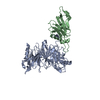 7mniC 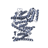 7mnjC  7mnkC 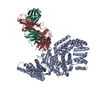 7mnlC 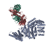 7mnmC 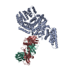 7mnnC  7mnoC 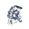 7mnpC 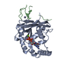 7mnqC 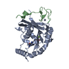 7mnrC  7mnsC  7mntC  7mnuC  7mnvC 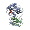 7mnwC 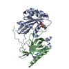 7mnxC 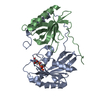 7mnyC 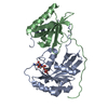 7mnzC 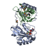 7mo0C 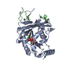 7mo1C  7mo2C 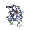 7mo3C 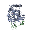 7mo4C 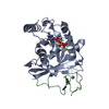 7mo5C 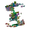 7tbmC M: map data used to model this data C: citing same article ( |
|---|


-Links
 PDBj
PDBj
- Assembly
| Deposited unit | 
|
|---|---|
| 1 | x 8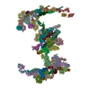
|
-Components
+Protein , 33 types, 102 molecules A1A3A2A4A5A6D1D2D3D4D5D6D7F1F2G1G2I1I2I3I4I5J1J2J3J4J5M1M2M3...
-Protein/peptide , 11 types, 39 molecules B1B2B3B4B5B6C1C2C3C4C5C6E1E2E3E4E5E6E7H1H2K1K2K3K4K5L1L2L3L4...
| #4: Protein/peptide | Mass: 1612.008 Da / Num. of mol.: 6 / Source method: isolated from a natural source Details: Crystal structure of the Chaetomium thermophilum NUP53 R3 ortholog component of the Nup170-Nup53 heterodimer structure (PDB ID 5HAX). To remain faithful to experimentally determined ...Details: Crystal structure of the Chaetomium thermophilum NUP53 R3 ortholog component of the Nup170-Nup53 heterodimer structure (PDB ID 5HAX). To remain faithful to experimentally determined structures, we opted to interpret the cryo-ET map of the Homo sapiens NPC with ortholog crystal structures. Source: (natural) Homo sapiens (human)#5: Protein/peptide | Mass: 2258.661 Da / Num. of mol.: 6 / Source method: isolated from a natural source Details: Crystal structure of the Chaetomium thermophilum NUP98 R3 ortholog component of the Nup170-Nup145N heterodimer structure (PDB ID 5HB0). To remain faithful to experimentally determined ...Details: Crystal structure of the Chaetomium thermophilum NUP98 R3 ortholog component of the Nup170-Nup145N heterodimer structure (PDB ID 5HB0). To remain faithful to experimentally determined structures, we opted to interpret the cryo-ET map of the Homo sapiens NPC with ortholog crystal structures. Source: (natural) Homo sapiens (human)#7: Protein/peptide | Mass: 903.057 Da / Num. of mol.: 7 / Source method: isolated from a natural source Details: Crystal structure of the Homo sapiens NUP53 R2 component of the NUP93-NUP53 heterodimer crystal structure (PDB ID 7MW1) used to interpret the cryo-ET map of the Homo sapiens NPC. Source: (natural) Homo sapiens (human)#10: Protein/peptide | Mass: 1348.416 Da / Num. of mol.: 2 / Source method: isolated from a natural source Details: Single particle cryo-EM structure of the Chaetomium thermophilum NUP98 R2 ortholog component of the Nup188-Nic96-Nup145N heterotrimer structure (PDB ID 7MVZ). To remain faithful to ...Details: Single particle cryo-EM structure of the Chaetomium thermophilum NUP98 R2 ortholog component of the Nup188-Nic96-Nup145N heterotrimer structure (PDB ID 7MVZ). To remain faithful to experimentally determined structures, we opted to interpret the cryo-ET map of the Homo sapiens NPC with ortholog single particle cryo-EM structures. Source: (natural) Homo sapiens (human)#13: Protein/peptide | Mass: 1062.345 Da / Num. of mol.: 5 / Source method: isolated from a natural source Details: Single particle cryo-EM structure of the Chaetomium thermophilum NUP98 R1 ortholog component of the Nup192-Nic96-Nup53-Nup145N heterotetramer structure (PDB ID 7MVV). To remain faithful to ...Details: Single particle cryo-EM structure of the Chaetomium thermophilum NUP98 R1 ortholog component of the Nup192-Nic96-Nup53-Nup145N heterotetramer structure (PDB ID 7MVV). To remain faithful to experimentally determined structures, we opted to interpret the cryo-ET map of the Homo sapiens NPC with ortholog single particle cryo-EM structures. Source: (natural) Homo sapiens (human)#14: Protein/peptide | Mass: 222.241 Da / Num. of mol.: 5 / Source method: isolated from a natural source Details: Single particle cryo-EM structure of the Chaetomium thermophilum NUP53 R1 ortholog component of the Nup192-Nic96-Nup53-Nup145N heterotetramer structure (PDB ID 7MVV). To remain faithful to ...Details: Single particle cryo-EM structure of the Chaetomium thermophilum NUP53 R1 ortholog component of the Nup192-Nic96-Nup53-Nup145N heterotetramer structure (PDB ID 7MVV). To remain faithful to experimentally determined structures, we opted to interpret the cryo-ET map of the Homo sapiens NPC with ortholog single particle cryo-EM structures. Source: (natural) Homo sapiens (human)#20: Protein/peptide | Mass: 4441.164 Da / Num. of mol.: 4 / Source method: isolated from a natural source Details: Crystal structure of the Chaetomium thermophilum NUP93 R1 ortholog component of the Nup49-Nup57-Nsp1-Nic96 heterotetramer structure (PDB ID 5CWS). To remain faithful to experimentally ...Details: Crystal structure of the Chaetomium thermophilum NUP93 R1 ortholog component of the Nup49-Nup57-Nsp1-Nic96 heterotetramer structure (PDB ID 5CWS). To remain faithful to experimentally determined structures, we opted to interpret the cryo-ET map of the Homo sapiens NPC with ortholog crystal structures. Source: (natural) Homo sapiens (human)#34: Protein/peptide | | Mass: 5141.046 Da / Num. of mol.: 1 / Source method: isolated from a natural source Details: Crystal structure of the Homo sapiens NUP42 GBM component of the GLE1-NUP42 heterodimer structure (PDB ID 6B4F) used to interpret the cryo-ET map of the Homo sapiens NPC. Source: (natural) Homo sapiens (human)#40: Protein/peptide | | Mass: 4416.920 Da / Num. of mol.: 1 / Source method: isolated from a natural source Details: Crystal structure of Xenopus laevis NUP62 CCS2 ortholog component of the channel nucleoporin heterotrimer (CNT; NUP62-NUP54-NUP58) structure (PDB ID 5C3L). To remain faithful to ...Details: Crystal structure of Xenopus laevis NUP62 CCS2 ortholog component of the channel nucleoporin heterotrimer (CNT; NUP62-NUP54-NUP58) structure (PDB ID 5C3L). To remain faithful to experimentally determined structures, we opted to interpret the cryo-ET map of the Homo sapiens NPC with ortholog crystal structures. Source: (natural) Homo sapiens (human)#42: Protein/peptide | | Mass: 3337.105 Da / Num. of mol.: 1 / Source method: isolated from a natural source Details: Poly-alanine model of the NUP214 CCS2 based on the Xenopus laevis and Chaetomium thermophilum channel nucleoporin heterotrimer (CNT; NUP62-NUP54-NUP58) crystal structures (PDB ID 5C3L and 5CWS). Source: (natural) Homo sapiens (human)#44: Protein/peptide | | Mass: 1720.111 Da / Num. of mol.: 1 / Source method: isolated from a natural source Details: Poly-alanine model of the NUP88 CCS2 based on the Xenopus laevis and Chaetomium thermophilum channel nucleoporin heterotrimer (CNT; NUP62-NUP54-NUP58) crystal structures (PDB ID 5C3L and 5CWS). Source: (natural) Homo sapiens (human) |
|---|
-Details
| Has ligand of interest | N |
|---|---|
| Has protein modification | Y |
-Experimental details
-Experiment
| Experiment | Method: ELECTRON MICROSCOPY |
|---|---|
| EM experiment | Aggregation state: PARTICLE / 3D reconstruction method: subtomogram averaging |
- Sample preparation
| Component | Name: Nuclear pore complex from purified HeLa cell nuclear envelopes. Type: COMPLEX / Entity ID: #1-#2, #4-#44 / Source: NATURAL |
|---|---|
| Source (natural) | Organism: Homo sapiens (human) |
| Buffer solution | pH: 7.5 |
| Specimen | Embedding applied: NO / Shadowing applied: NO / Staining applied: NO / Vitrification applied: YES |
| Vitrification | Cryogen name: ETHANE |
- Electron microscopy imaging
| Experimental equipment |  Model: Titan Krios / Image courtesy: FEI Company |
|---|---|
| Microscopy | Model: FEI TITAN KRIOS |
| Electron gun | Electron source:  FIELD EMISSION GUN / Accelerating voltage: 300 kV / Illumination mode: FLOOD BEAM FIELD EMISSION GUN / Accelerating voltage: 300 kV / Illumination mode: FLOOD BEAM |
| Electron lens | Mode: BRIGHT FIELD / Nominal defocus max: 4000 nm / Nominal defocus min: 1900 nm |
| Image recording | Electron dose: 4 e/Å2 / Avg electron dose per subtomogram: 110 e/Å2 / Film or detector model: GATAN K2 SUMMIT (4k x 4k) |
- Processing
| CTF correction | Type: PHASE FLIPPING AND AMPLITUDE CORRECTION |
|---|---|
| 3D reconstruction | Resolution: 23 Å / Resolution method: FSC 0.143 CUT-OFF / Num. of particles: 17368 / Symmetry type: POINT |
| EM volume selection | Num. of tomograms: 170 / Num. of volumes extracted: 17368 |
| Atomic model building | Details: Authors state that the clashes between nucleoporin structures result from docking nucleoporin structures from different species into low-resolution cryo-ET maps of intact NPCs without flexible fitting. |
