 Movie
Movie Controller

[English] 日本語
 Yorodumi
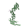
- PDB-7tbi: Composite structure of the S. cerevisiae nuclear pore complex (NPC) -
+ Open data

- Basic information
| Entry | Database: PDB / ID: 7tbi | |||||||||||||||
|---|---|---|---|---|---|---|---|---|---|---|---|---|---|---|---|---|
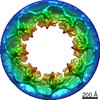
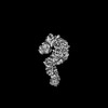
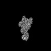
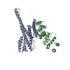
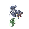
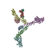
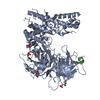
| Title | Composite structure of the S. cerevisiae nuclear pore complex (NPC) | |||||||||||||||
 Components Components |
| |||||||||||||||
 Keywords Keywords | TRANSPORT PROTEIN / nuclear pore complex / nucleocytoplasmic transport / alpha-helical solenoid / nuclear pore | |||||||||||||||
| Biological species |  | |||||||||||||||
| Method | ELECTRON MICROSCOPY / subtomogram averaging / cryo EM / Resolution: 25 Å | |||||||||||||||
 Authors Authors | Petrovic, S. / Samanta, D. / Perriches, T. / Bley, C.J. / Thierbach, K. / Brown, B. / Nie, S. / Mobbs, G.W. / Stevens, T.A. / Liu, X. ...Petrovic, S. / Samanta, D. / Perriches, T. / Bley, C.J. / Thierbach, K. / Brown, B. / Nie, S. / Mobbs, G.W. / Stevens, T.A. / Liu, X. / Tomaleri, G.P. / Schaus, L. / Hoelz, A. | |||||||||||||||
| Funding support |  United States, 4items United States, 4items
| |||||||||||||||
 Citation Citation | Journal: Nature / Year: 2020 Title: In-cell architecture of the nuclear pore and snapshots of its turnover. Authors: Matteo Allegretti / Christian E Zimmerli / Vasileios Rantos / Florian Wilfling / Paolo Ronchi / Herman K H Fung / Chia-Wei Lee / Wim Hagen / Beata Turoňová / Kai Karius / Mandy Börmel / ...Authors: Matteo Allegretti / Christian E Zimmerli / Vasileios Rantos / Florian Wilfling / Paolo Ronchi / Herman K H Fung / Chia-Wei Lee / Wim Hagen / Beata Turoňová / Kai Karius / Mandy Börmel / Xiaojie Zhang / Christoph W Müller / Yannick Schwab / Julia Mahamid / Boris Pfander / Jan Kosinski / Martin Beck /  Abstract: Nuclear pore complexes (NPCs) fuse the inner and outer membranes of the nuclear envelope. They comprise hundreds of nucleoporins (Nups) that assemble into multiple subcomplexes and form large central ...Nuclear pore complexes (NPCs) fuse the inner and outer membranes of the nuclear envelope. They comprise hundreds of nucleoporins (Nups) that assemble into multiple subcomplexes and form large central channels for nucleocytoplasmic exchange. How this architecture facilitates messenger RNA export, NPC biogenesis and turnover remains poorly understood. Here we combine in situ structural biology and integrative modelling with correlative light and electron microscopy and molecular perturbation to structurally analyse NPCs in intact Saccharomyces cerevisiae cells within the context of nuclear envelope remodelling. We find an in situ conformation and configuration of the Nup subcomplexes that was unexpected from the results of previous in vitro analyses. The configuration of the Nup159 complex appears critical to spatially accommodate its function as an mRNA export platform, and as a mediator of NPC turnover. The omega-shaped nuclear envelope herniae that accumulate in nup116Δ cells conceal partially assembled NPCs lacking multiple subcomplexes, including the Nup159 complex. Under conditions of starvation, herniae of a second type are formed that cytoplasmically expose NPCs. These results point to a model of NPC turnover in which NPC-containing vesicles bud off from the nuclear envelope before degradation by the autophagy machinery. Our study emphasizes the importance of investigating the structure-function relationship of macromolecular complexes in their cellular context. | |||||||||||||||
| History |
| |||||||||||||||
| Remark 0 | THIS ENTRY 7TBI REFLECTS AN ALTERNATIVE MODELING OF THE ORIGINAL DATA IN EMD-10198, DETERMINED BY M. ...THIS ENTRY 7TBI REFLECTS AN ALTERNATIVE MODELING OF THE ORIGINAL DATA IN EMD-10198, DETERMINED BY M.Beck,M.Allegretti |
- Structure visualization
| Structure viewer | Molecule:  MolmilJmol/JSmol MolmilJmol/JSmol |
|---|
- Downloads & links
-Download
| PDBx/mmCIF format | 7tbi.cif.gz | 5.1 MB | Display | PDBx/mmCIF format |
|---|---|---|---|---|
| PDB format | pdb7tbi.ent.gz | Display | PDB format | |
| PDBx/mmJSON format | 7tbi.json.gz | Tree view | PDBx/mmJSON format | |
| Others |  Other downloads Other downloads |
-Validation report
| Arichive directory | https://data.pdbj.org/pub/pdb/validation_reports/tb/7tbiftp://data.pdbj.org/pub/pdb/validation_reports/tb/7tbi | HTTPS FTP |
|---|
-Related structure data
| Related structure data |  7mvtC  7mvuC  7mvvC  7mvwC  7mvxC  7mvyC  7mvzC  7mw0C  7mw1C  7tbjC 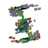 7tbkC C: citing same article ( M: map data used to model this data |
|---|---|
| EM raw data | EMPIAR-10466 (Title: Tilt-series from cryo-lamellae of WT S. cerevisiae cells Data size: 605.7 Data #1: Raw tilt series from S. cerevisiae WT cells containing nuclear pore complexes (each tilt is an aligned average of ~15 frames by serial em) [tilt series] Data #2: Tilt series of WT cerevisiae cells after cleaning bad tilts and dose filtering [tilt series]) |


-Links
 PDBj
PDBj
- Assembly
| Deposited unit | 
|
|---|---|
| 1 | x 8
|
-Components
+Protein , 22 types, 52 molecules A2A3A4A1D1D2D3D4F1F2G1G2I1I2J1J2M1M2M3M4N1N2N3N4O1O2O3O4Q1Q2...
-Protein/peptide , 4 types, 14 molecules B1B2B3B4E1E2E3E4L1L2P1P2P3P4
| #2: Protein/peptide | Mass: 1612.008 Da / Num. of mol.: 4 / Source method: isolated from a natural source Details: Crystal structure of the Chaetomium thermophilum Nup53/Nup59 R3 ortholog component of the the Nup170-Nup53 heterodimer structure (PDB ID 5HAX). To remain faithful to experimentally ...Details: Crystal structure of the Chaetomium thermophilum Nup53/Nup59 R3 ortholog component of the the Nup170-Nup53 heterodimer structure (PDB ID 5HAX). To remain faithful to experimentally determined structures, we opted to interpret the in situ cryo-ET map of the S. cerevisiae NPC with C. thermophilum crystal structures. Source: (natural) #5: Protein/peptide | Mass: 1949.145 Da / Num. of mol.: 4 / Source method: isolated from a natural source Details: Crystal structure of the Chaetomium thermophilum Nup53/Nup59 R2 ortholog component of the the Nic96-Nup53 heterodimer crystal structure (PDB ID 5HB3). To remain faithful to experimentally ...Details: Crystal structure of the Chaetomium thermophilum Nup53/Nup59 R2 ortholog component of the the Nic96-Nup53 heterodimer crystal structure (PDB ID 5HB3). To remain faithful to experimentally determined structures, we opted to interpret the in situ cryo-ET map of the S. cerevisiae NPC with C. thermophilum crystal structures. Source: (natural) #12: Protein/peptide | Mass: 222.241 Da / Num. of mol.: 2 / Source method: isolated from a natural source Details: Single particle cryo-EM structure of the Chaetomium thermophilum Nup53/Nup59 R1 ortholog component of the the Nup192-Nic96-Nup53-Nup145N heterotetramer structure (PDB ID 7MVV). To remain ...Details: Single particle cryo-EM structure of the Chaetomium thermophilum Nup53/Nup59 R1 ortholog component of the the Nup192-Nic96-Nup53-Nup145N heterotetramer structure (PDB ID 7MVV). To remain faithful to experimentally determined structures, we opted to interpret the in situ cryo-ET map of the S. cerevisiae NPC with C. thermophilum single particle cryo-EM structures. Source: (natural) #16: Protein/peptide | Mass: 4441.164 Da / Num. of mol.: 4 / Source method: isolated from a natural source Details: Crystal structure of Chaetomium thermophilum Nic96 R1 ortholog component of the Nup49-Nup57-Nsp1-Nic96 heterotetramer structure (PDB ID 5CWS). To remain faithful to experimentally determined ...Details: Crystal structure of Chaetomium thermophilum Nic96 R1 ortholog component of the Nup49-Nup57-Nsp1-Nic96 heterotetramer structure (PDB ID 5CWS). To remain faithful to experimentally determined structures, we opted to interpret the in situ cryo-ET map of the S. cerevisiae NPC with C. thermophilum crystal structures. Source: (natural) |
|---|
-Nup145N/Nup100/Nup116 ... , 3 types, 8 molecules C2C3C1C4H1H2K1K2
| #3: Protein/peptide | Mass: 1991.335 Da / Num. of mol.: 4 / Source method: isolated from a natural source Details: Crystal structure of the Chaetomium thermophilum Nup145N/Nup100/Nup116 R3 ortholog component of the the Nup170-Nup145N heterodimer structure (PDB ID 5HB0). To remain faithful to ...Details: Crystal structure of the Chaetomium thermophilum Nup145N/Nup100/Nup116 R3 ortholog component of the the Nup170-Nup145N heterodimer structure (PDB ID 5HB0). To remain faithful to experimentally determined structures, we opted to interpret the in situ cryo-ET map of the S. cerevisiae NPC with C. thermophilum crystal structures. Source: (natural) #8: Protein/peptide | Mass: 1348.416 Da / Num. of mol.: 2 / Source method: isolated from a natural source Details: Single particle cryo-EM structure of the Chaetomium thermophilum Nup145N/Nup100/Nup116 R2 ortholog component of the the Nup188-Nic96-Nup145N heterotrimer structure (PDB ID 7MVZ). To remain ...Details: Single particle cryo-EM structure of the Chaetomium thermophilum Nup145N/Nup100/Nup116 R2 ortholog component of the the Nup188-Nic96-Nup145N heterotrimer structure (PDB ID 7MVZ). To remain faithful to experimentally determined structures, we opted to interpret the in situ cryo-ET map of the S. cerevisiae NPC with C. thermophilum single particle cryo-EM structures. Source: (natural) #11: Protein/peptide | Mass: 1062.345 Da / Num. of mol.: 2 / Source method: isolated from a natural source Details: Single particle cryo-EM structure of the Chaetomium thermophilum Nup145N/Nup100/Nup116 R1 ortholog component of the the Nup192-Nic96-Nup53-Nup145N heterotetramer structure (PDB ID 7MVV). To ...Details: Single particle cryo-EM structure of the Chaetomium thermophilum Nup145N/Nup100/Nup116 R1 ortholog component of the the Nup192-Nic96-Nup53-Nup145N heterotetramer structure (PDB ID 7MVV). To remain faithful to experimentally determined structures, we opted to interpret the in situ cryo-ET map of the S. cerevisiae NPC with C. thermophilum single particle cryo-EM structures. Source: (natural) |
|---|
-Details
| Has ligand of interest | N |
|---|---|
| Has protein modification | Y |
-Experimental details
-Experiment
| Experiment | Method: ELECTRON MICROSCOPY |
|---|---|
| EM experiment | Aggregation state: CELL / 3D reconstruction method: subtomogram averaging |
- Sample preparation
| Component |
| ||||||||||||||||||||||||||||||
|---|---|---|---|---|---|---|---|---|---|---|---|---|---|---|---|---|---|---|---|---|---|---|---|---|---|---|---|---|---|---|---|
| Source (natural) |
| ||||||||||||||||||||||||||||||
| Buffer solution | pH: 7.5 | ||||||||||||||||||||||||||||||
| Specimen | Embedding applied: NO / Shadowing applied: NO / Staining applied: NO / Vitrification applied: YES | ||||||||||||||||||||||||||||||
| Vitrification | Cryogen name: ETHANE |
- Electron microscopy imaging
| Experimental equipment |  Model: Titan Krios / Image courtesy: FEI Company |
|---|---|
| Microscopy | Model: FEI TITAN KRIOS |
| Electron gun | Electron source:  FIELD EMISSION GUN / Accelerating voltage: 300 kV / Illumination mode: FLOOD BEAM FIELD EMISSION GUN / Accelerating voltage: 300 kV / Illumination mode: FLOOD BEAM |
| Electron lens | Mode: BRIGHT FIELD / Nominal defocus max: 5000 nm / Nominal defocus min: 2000 nm |
| Image recording | Electron dose: 4 e/Å2 / Avg electron dose per subtomogram: 140 e/Å2 / Film or detector model: GATAN K2 SUMMIT (4k x 4k) |
- Processing
| CTF correction | Type: PHASE FLIPPING AND AMPLITUDE CORRECTION |
|---|---|
| 3D reconstruction | Resolution: 25 Å / Resolution method: FSC 0.143 CUT-OFF / Num. of particles: 4000 / Symmetry type: POINT |
| EM volume selection | Num. of tomograms: 240 / Num. of volumes extracted: 4000 |
| Atomic model building | Details: Authors state that the clashes between nucleoporin structures result from docking nucleoporin structures from different species into low-resolution cryo-ET maps of intact NPCs without flexible fitting. |
