 Movie
Movie Controller

[English] 日本語
 Yorodumi
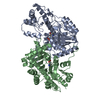
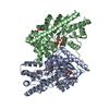
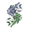
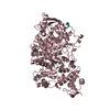
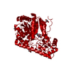
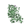
- PDB-3pbp: Structure of the yeast heterotrimeric Nup82-Nup159-Nup116 nucleop... -
+ Open data

- Basic information
| Entry | Database: PDB / ID: 3pbp | ||||||
|---|---|---|---|---|---|---|---|
| Title | Structure of the yeast heterotrimeric Nup82-Nup159-Nup116 nucleoporin complex | ||||||
 Components Components |
| ||||||
 Keywords Keywords | TRANSPORT PROTEIN / STRUCTURAL PROTEIN / beta-propeller / nucleoporin / mRNA export / mRNP remodelling / NUCLEOCYTOPLASMIC Transport / PROTEIN TRANSPORT / TRANSLOCATION / TRANSPORT / autoproteolysis / Fusion protein / PROTOONCOGENE / ONCOPROTEIN / Protein COMPLEX / Nucleus / Nuclear Envelope / Nuclear Pore Complex | ||||||
| Function / homology |  Function and homology information Function and homology informationnuclear pore linkers / nuclear pore localization / adenyl-nucleotide exchange factor activity / nuclear pore central transport channel / Regulation of Glucokinase by Glucokinase Regulatory Protein / telomere tethering at nuclear periphery / : / nuclear pore organization / nuclear pore cytoplasmic filaments / Regulation of HSF1-mediated heat shock response ...nuclear pore linkers / nuclear pore localization / adenyl-nucleotide exchange factor activity / nuclear pore central transport channel / Regulation of Glucokinase by Glucokinase Regulatory Protein / telomere tethering at nuclear periphery / : / nuclear pore organization / nuclear pore cytoplasmic filaments / Regulation of HSF1-mediated heat shock response / post-transcriptional tethering of RNA polymerase II gene DNA at nuclear periphery / tRNA export from nucleus / SUMOylation of SUMOylation proteins / structural constituent of nuclear pore / SUMOylation of RNA binding proteins / RNA export from nucleus / SUMOylation of chromatin organization proteins / nucleocytoplasmic transport / nuclear localization sequence binding / NLS-bearing protein import into nucleus / poly(A)+ mRNA export from nucleus / nuclear pore / ribosomal large subunit export from nucleus / mRNA export from nucleus / ribosomal small subunit export from nucleus / protein export from nucleus / protein import into nucleus / transcription corepressor activity / nuclear envelope / ATPase binding / nuclear membrane / RNA binding / identical protein binding / nucleus / cytosol Similarity search - Function | ||||||
| Biological species |  | ||||||
| Method |  X-RAY DIFFRACTION / SYNCHROTRON / SAD / Resolution: 2.6 Å X-RAY DIFFRACTION / SYNCHROTRON / SAD / Resolution: 2.6 Å | ||||||
 Authors Authors | Debler, E.W. / Hoelz, A. | ||||||
 Citation Citation | Journal: Proc.Natl.Acad.Sci.USA / Year: 2011 Title: Structural and functional analysis of an essential nucleoporin heterotrimer on the cytoplasmic face of the nuclear pore complex. Authors: Yoshida, K. / Seo, H.S. / Debler, E.W. / Blobel, G. / Hoelz, A. | ||||||
| History |
|
- Structure visualization
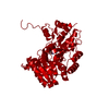
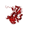
| Structure viewer | Molecule: MolmilJmol/JSmol |
|---|
- Downloads & links
-Download
| PDBx/mmCIF format | 3pbp.cif.gz | 483.2 KB | Display | PDBx/mmCIF format |
|---|---|---|---|---|
| PDB format | pdb3pbp.ent.gz | 397.6 KB | Display | PDB format |
| PDBx/mmJSON format | 3pbp.json.gz | Tree view | PDBx/mmJSON format | |
| Others |  Other downloads Other downloads |
-Validation report
| Arichive directory | https://data.pdbj.org/pub/pdb/validation_reports/pb/3pbpftp://data.pdbj.org/pub/pdb/validation_reports/pb/3pbp | HTTPS FTP |
|---|
-Related structure data
| Similar structure data |
|---|

-Links
 PDBj
PDBj

- Assembly
| Deposited unit | 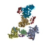
| ||||||||
|---|---|---|---|---|---|---|---|---|---|
| 1 | 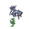
| ||||||||
| 2 | 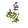
| ||||||||
| 3 | 
| ||||||||
| 4 | 
| ||||||||
| Unit cell |
|
-Components
| #1: Protein | Mass: 51982.461 Da / Num. of mol.: 4 / Fragment: N-terminal domain (NTD), UNP residues 1-452 / Mutation: C396S Source method: isolated from a genetically manipulated source Source: (gene. exp.) Gene: NUP82, YJL061W, J1135, HRB187 / Production host:  #2: Protein | Mass: 16832.180 Da / Num. of mol.: 4 / Fragment: C-terminal domain (CTD), UNP residues 967-1113 Source method: isolated from a genetically manipulated source Source: (gene. exp.) Gene: NUP116, NSP116, YMR047C, YM9532.12C / Production host: #3: Protein/peptide | Mass: 4167.491 Da / Num. of mol.: 4 / Fragment: Tail, UNP residues 1425-1460 Source method: isolated from a genetically manipulated source Source: (gene. exp.) Gene: NUP159, NUP158, RAT7, YIL115C / Production host: #4: Chemical | ChemComp-PGE / |   Mass: 150.173 Da / Num. of mol.: 1 / Source method: obtained synthetically / Formula: C6H14O4 Mass: 150.173 Da / Num. of mol.: 1 / Source method: obtained synthetically / Formula: C6H14O4Has protein modification | Y | |
|---|
-Experimental details
-Experiment
| Experiment | Method: X-RAY DIFFRACTION / Number of used crystals: 1 |
|---|
- Sample preparation
| Crystal | Density Matthews: 2.65 Å3/Da / Density % sol: 53.52 % |
|---|---|
| Crystal grow | Temperature: 298 K / Method: vapor diffusion, sitting drop / pH: 6.6 Details: PEG 400, sodium cacodylate, lithium sulfate, 2,5-hexanediol, pH 6.6, VAPOR DIFFUSION, SITTING DROP, temperature 298K |
-Data collection
| Diffraction | Mean temperature: 100 K |
|---|---|
| Diffraction source | Source: SYNCHROTRON / Site: APS  / Beamline: 23-ID-B / Wavelength: 0.9794 Å / Beamline: 23-ID-B / Wavelength: 0.9794 Å |
| Detector | Type: MARMOSAIC 300 mm CCD / Detector: CCD / Date: Feb 9, 2009 |
| Radiation | Monochromator: double crystal / Protocol: SINGLE WAVELENGTH / Monochromatic (M) / Laue (L): M / Scattering type: x-ray |
| Radiation wavelength | Wavelength: 0.9794 Å / Relative weight: 1 |
| Reflection | Resolution: 2.6→50 Å / Num. all: 184127 / Num. obs: 181917 / % possible obs: 98.8 % / Observed criterion σ(I): -3 / Redundancy: 2 % / Rsym value: 0.097 / Net I/σ(I): 11 |
| Reflection shell | Resolution: 2.6→2.69 Å / Redundancy: 2 % / Mean I/σ(I) obs: 3 / Num. unique all: 17910 / Rsym value: 0.473 / % possible all: 98.3 |
- Processing
| Software |
| ||||||||||||||||||||
|---|---|---|---|---|---|---|---|---|---|---|---|---|---|---|---|---|---|---|---|---|---|
| Refinement | Method to determine structure: SAD / Resolution: 2.6→50 Å / σ(F): 0 / Stereochemistry target values: Engh & Huber
| ||||||||||||||||||||
| Refinement step | Cycle: LAST / Resolution: 2.6→50 Å
| ||||||||||||||||||||
| Refine LS restraints |
|
