Journal: Nature / Year: 2020 Title: In-cell architecture of the nuclear pore and snapshots of its turnover. Authors: Matteo Allegretti / Christian E Zimmerli / Vasileios Rantos / Florian Wilfling / Paolo Ronchi / Herman K H Fung / Chia-Wei Lee / Wim Hagen / Beata Turoňová / Kai Karius / Mandy Börmel / ...Authors: Matteo Allegretti / Christian E Zimmerli / Vasileios Rantos / Florian Wilfling / Paolo Ronchi / Herman K H Fung / Chia-Wei Lee / Wim Hagen / Beata Turoňová / Kai Karius / Mandy Börmel / Xiaojie Zhang / Christoph W Müller / Yannick Schwab / Julia Mahamid / Boris Pfander / Jan Kosinski / Martin Beck / Abstract: Nuclear pore complexes (NPCs) fuse the inner and outer membranes of the nuclear envelope. They comprise hundreds of nucleoporins (Nups) that assemble into multiple subcomplexes and form large central ...Nuclear pore complexes (NPCs) fuse the inner and outer membranes of the nuclear envelope. They comprise hundreds of nucleoporins (Nups) that assemble into multiple subcomplexes and form large central channels for nucleocytoplasmic exchange. How this architecture facilitates messenger RNA export, NPC biogenesis and turnover remains poorly understood. Here we combine in situ structural biology and integrative modelling with correlative light and electron microscopy and molecular perturbation to structurally analyse NPCs in intact Saccharomyces cerevisiae cells within the context of nuclear envelope remodelling. We find an in situ conformation and configuration of the Nup subcomplexes that was unexpected from the results of previous in vitro analyses. The configuration of the Nup159 complex appears critical to spatially accommodate its function as an mRNA export platform, and as a mediator of NPC turnover. The omega-shaped nuclear envelope herniae that accumulate in nup116Δ cells conceal partially assembled NPCs lacking multiple subcomplexes, including the Nup159 complex. Under conditions of starvation, herniae of a second type are formed that cytoplasmically expose NPCs. These results point to a model of NPC turnover in which NPC-containing vesicles bud off from the nuclear envelope before degradation by the autophagy machinery. Our study emphasizes the importance of investigating the structure-function relationship of macromolecular complexes in their cellular context.
History
Deposition
Aug 7, 2019
-
Header (metadata) release
Mar 18, 2020
-
Map release
Aug 26, 2020
-
Update
Feb 10, 2021
-
Current status
Feb 10, 2021
Processing site: PDBe / Status: Released
-
Structure visualization
Movie
Surface view with section colored by density value
EMPIAR-10466 (Title: Tilt-series from cryo-lamellae of WT S. cerevisiae cells Data size: 605.7 Data #1: Raw tilt series from S. cerevisiae WT cells containing nuclear pore complexes (each tilt is an aligned average of ~15 frames by serial em) [tilt series] Data #2: Tilt series of WT cerevisiae cells after cleaning bad tilts and dose filtering [tilt series])
In the structure databanks used in Yorodumi, some data are registered as the other names, "COVID-19 virus" and "2019-nCoV". Here are the details of the virus and the list of structure data.
Jan 31, 2019. EMDB accession codes are about to change! (news from PDBe EMDB page)
EMDB accession codes are about to change! (news from PDBe EMDB page)
The allocation of 4 digits for EMDB accession codes will soon come to an end. Whilst these codes will remain in use, new EMDB accession codes will include an additional digit and will expand incrementally as the available range of codes is exhausted. The current 4-digit format prefixed with “EMD-” (i.e. EMD-XXXX) will advance to a 5-digit format (i.e. EMD-XXXXX), and so on. It is currently estimated that the 4-digit codes will be depleted around Spring 2019, at which point the 5-digit format will come into force.
The EM Navigator/Yorodumi systems omit the EMD- prefix.
Related info.:Q: What is EMD? / ID/Accession-code notation in Yorodumi/EM Navigator
Yorodumi is a browser for structure data from EMDB, PDB, SASBDB, etc.
This page is also the successor to EM Navigator detail page, and also detail information page/front-end page for Omokage search.
The word "yorodu" (or yorozu) is an old Japanese word meaning "ten thousand". "mi" (miru) is to see.
Related info.:EMDB / PDB / SASBDB / Comparison of 3 databanks / Yorodumi Search / Aug 31, 2016. New EM Navigator & Yorodumi / Yorodumi Papers / Jmol/JSmol / Function and homology information / Changes in new EM Navigator and Yorodumi
 Movie
Movie Controller


 Open data
 Basic information
 Map data
 Sample
 Authors
 Citation
 Structure visualization
 Movie viewer
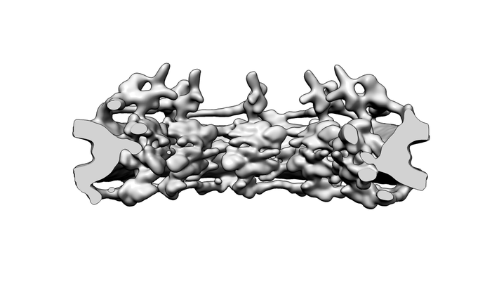
 Downloads & links
 emd_10198.png
 http://ftp.pdbj.org/pub/emdb/structures/EMD-10198


 Z (Sec.)
 Y (Row.)
 X (Col.)


 Sample components
 Processing
 Electron microscopy
 FIELD EMISSION GUN
