 Movie
Movie Controller

[English] 日本語
 Yorodumi
- PDB-7tbk: Composite structure of the dilated human nuclear pore complex (NP... -
+ Open data

- Basic information
| Entry | Database: PDB / ID: 7tbk | |||||||||||||||
|---|---|---|---|---|---|---|---|---|---|---|---|---|---|---|---|---|
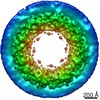
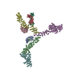
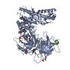
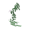
| Title | Composite structure of the dilated human nuclear pore complex (NPC) symmetric core generated with a 37A in situ cryo-ET map of CD4+ T cell NPC | |||||||||||||||
 Components Components |
| |||||||||||||||
 Keywords Keywords | TRANSPORT PROTEIN / nuclear pore complex / nucleocytoplasmic transport / alpha-helical solenoid / nuclear pore | |||||||||||||||
| Biological species |  Homo sapiens (human) Homo sapiens (human) | |||||||||||||||
| Method | ELECTRON MICROSCOPY / subtomogram averaging / cryo EM / Resolution: 37 Å | |||||||||||||||
 Authors Authors | Petrovic, S. / Samanta, D. / Perriches, T. / Bley, C.J. / Thierbach, K. / Brown, B. / Nie, S. / Mobbs, G.W. / Stevens, T.A. / Liu, X. ...Petrovic, S. / Samanta, D. / Perriches, T. / Bley, C.J. / Thierbach, K. / Brown, B. / Nie, S. / Mobbs, G.W. / Stevens, T.A. / Liu, X. / Tomaleri, G.P. / Schaus, L. / Hoelz, A. | |||||||||||||||
| Funding support |  United States, 4items United States, 4items
| |||||||||||||||
 Citation Citation | Journal: Cell / Year: 2021 Title: Cone-shaped HIV-1 capsids are transported through intact nuclear pores. Authors: Vojtech Zila / Erica Margiotta / Beata Turoňová / Thorsten G Müller / Christian E Zimmerli / Simone Mattei / Matteo Allegretti / Kathleen Börner / Jona Rada / Barbara Müller / Marina ...Authors: Vojtech Zila / Erica Margiotta / Beata Turoňová / Thorsten G Müller / Christian E Zimmerli / Simone Mattei / Matteo Allegretti / Kathleen Börner / Jona Rada / Barbara Müller / Marina Lusic / Hans-Georg Kräusslich / Martin Beck /  Abstract: Human immunodeficiency virus (HIV-1) remains a major health threat. Viral capsid uncoating and nuclear import of the viral genome are critical for productive infection. The size of the HIV-1 capsid ...Human immunodeficiency virus (HIV-1) remains a major health threat. Viral capsid uncoating and nuclear import of the viral genome are critical for productive infection. The size of the HIV-1 capsid is generally believed to exceed the diameter of the nuclear pore complex (NPC), indicating that capsid uncoating has to occur prior to nuclear import. Here, we combined correlative light and electron microscopy with subtomogram averaging to capture the structural status of reverse transcription-competent HIV-1 complexes in infected T cells. We demonstrated that the diameter of the NPC in cellulo is sufficient for the import of apparently intact, cone-shaped capsids. Subsequent to nuclear import, we detected disrupted and empty capsid fragments, indicating that uncoating of the replication complex occurs by breaking the capsid open, and not by disassembly into individual subunits. Our data directly visualize a key step in HIV-1 replication and enhance our mechanistic understanding of the viral life cycle. | |||||||||||||||
| History |
| |||||||||||||||
| Remark 0 | THIS ENTRY 7TBK REFLECTS AN ALTERNATIVE MODELING OF THE ORIGINAL DATA IN EMD-11967, DETERMINED BY E. ...THIS ENTRY 7TBK REFLECTS AN ALTERNATIVE MODELING OF THE ORIGINAL DATA IN EMD-11967, DETERMINED BY E.Margiotta,M.Beck |
- Structure visualization
| Structure viewer | Molecule:  MolmilJmol/JSmol MolmilJmol/JSmol |
|---|
- Downloads & links
-Download
| PDBx/mmCIF format | 7tbk.cif.gz | 8.4 MB | Display | PDBx/mmCIF format |
|---|---|---|---|---|
| PDB format | pdb7tbk.ent.gz | Display | PDB format | |
| PDBx/mmJSON format | 7tbk.json.gz | Tree view | PDBx/mmJSON format | |
| Others |  Other downloads Other downloads |
-Validation report
| Arichive directory | https://data.pdbj.org/pub/pdb/validation_reports/tb/7tbkftp://data.pdbj.org/pub/pdb/validation_reports/tb/7tbk | HTTPS FTP |
|---|
-Related structure data
| Related structure data | 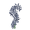 7mvtC  7mvuC  7mvvC  7mvwC 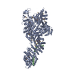 7mvxC  7mvyC  7mvzC 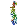 7mw0C 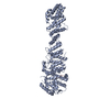 7mw1C  7tbiC  7tbjC M: map data used to model this data C: citing same article ( |
|---|


-Links
 PDBj
PDBj
- Assembly
| Deposited unit | 
|
|---|---|
| 1 | x 8
|
-Components
+Protein , 21 types, 79 molecules A1A3A2A4A5A6D1D2D3D4D5D6D7F1F2G1G2I1I2I3I4I5J1J2J3J4J5M1M2M3...
-Protein/peptide , 7 types, 35 molecules B1B2B3B4B5B6C1C2C3C4C5C6E1E2E3E4E5E6E7H1H2K1K2K3K4K5L1L2L3L4...
| #4: Protein/peptide | Mass: 1612.008 Da / Num. of mol.: 6 / Source method: isolated from a natural source Details: Crystal structure of the Chaetomium thermophilum NUP53 R3 ortholog component of the Nup170-Nup53 heterodimer structure (PDB ID 5HAX). To remain faithful to experimentally determined ...Details: Crystal structure of the Chaetomium thermophilum NUP53 R3 ortholog component of the Nup170-Nup53 heterodimer structure (PDB ID 5HAX). To remain faithful to experimentally determined structures, we opted to interpret the cryo-ET map of the Homo sapiens NPC with ortholog crystal structures. Source: (natural) Homo sapiens (human)#5: Protein/peptide | Mass: 2258.661 Da / Num. of mol.: 6 / Source method: isolated from a natural source Details: Crystal structure of the Chaetomium thermophilum NUP98 R3 ortholog component of the Nup170-Nup145N heterodimer structure (PDB ID 5HB0). To remain faithful to experimentally determined ...Details: Crystal structure of the Chaetomium thermophilum NUP98 R3 ortholog component of the Nup170-Nup145N heterodimer structure (PDB ID 5HB0). To remain faithful to experimentally determined structures, we opted to interpret the cryo-ET map of the Homo sapiens NPC with ortholog crystal structures. Source: (natural) Homo sapiens (human)#7: Protein/peptide | Mass: 903.057 Da / Num. of mol.: 7 / Source method: isolated from a natural source Details: Crystal structure of the Homo sapiens NUP53 R2 component of the NUP93-NUP53 heterodimer crystal structure (PDB ID 7MW1) used to interpret the cryo-ET map of the Homo sapiens NPC. Source: (natural) Homo sapiens (human)#10: Protein/peptide | Mass: 1348.416 Da / Num. of mol.: 2 / Source method: isolated from a natural source Details: Single particle cryo-EM structure of the Chaetomium thermophilum NUP98 R2 ortholog component of the Nup188-Nic96-Nup145N heterotrimer structure (PDB ID 7MVZ). To remain faithful to ...Details: Single particle cryo-EM structure of the Chaetomium thermophilum NUP98 R2 ortholog component of the Nup188-Nic96-Nup145N heterotrimer structure (PDB ID 7MVZ). To remain faithful to experimentally determined structures, we opted to interpret the cryo-ET map of the Homo sapiens NPC with ortholog single particle cryo-EM structures. Source: (natural) Homo sapiens (human)#13: Protein/peptide | Mass: 1062.345 Da / Num. of mol.: 5 / Source method: isolated from a natural source Details: Single particle cryo-EM structure of the Chaetomium thermophilum NUP98 R1 ortholog component of the Nup192-Nic96-Nup53-Nup145N heterotetramer structure (PDB ID 7MVV). To remain faithful to ...Details: Single particle cryo-EM structure of the Chaetomium thermophilum NUP98 R1 ortholog component of the Nup192-Nic96-Nup53-Nup145N heterotetramer structure (PDB ID 7MVV). To remain faithful to experimentally determined structures, we opted to interpret the cryo-ET map of the Homo sapiens NPC with ortholog single particle cryo-EM structures. Source: (natural) Homo sapiens (human)#14: Protein/peptide | Mass: 222.241 Da / Num. of mol.: 5 / Source method: isolated from a natural source Details: Single particle cryo-EM structure of the Chaetomium thermophilum NUP53 R1 ortholog component of the Nup192-Nic96-Nup53-Nup145N heterotetramer structure (PDB ID 7MVV). To remain faithful to ...Details: Single particle cryo-EM structure of the Chaetomium thermophilum NUP53 R1 ortholog component of the Nup192-Nic96-Nup53-Nup145N heterotetramer structure (PDB ID 7MVV). To remain faithful to experimentally determined structures, we opted to interpret the cryo-ET map of the Homo sapiens NPC with ortholog single particle cryo-EM structures. Source: (natural) Homo sapiens (human)#20: Protein/peptide | Mass: 4441.164 Da / Num. of mol.: 4 / Source method: isolated from a natural source Details: Crystal structure of the Chaetomium thermophilum NUP93 R1 ortholog component of the Nup49-Nup57-Nsp1-Nic96 heterotetramer structure (PDB ID 5CWS). To remain faithful to experimentally ...Details: Crystal structure of the Chaetomium thermophilum NUP93 R1 ortholog component of the Nup49-Nup57-Nsp1-Nic96 heterotetramer structure (PDB ID 5CWS). To remain faithful to experimentally determined structures, we opted to interpret the cryo-ET map of the Homo sapiens NPC with ortholog crystal structures. Source: (natural) Homo sapiens (human) |
|---|
-NUP54 Ferrodoxin-like ... , 2 types, 8 molecules P1P2P3P4Q1Q2Q3Q4
| #18: Protein | Mass: 12745.392 Da / Num. of mol.: 4 / Source method: isolated from a natural source Details: Crystal structure of Xenopus laevis NUP54 Ferrodoxin-like domain ortholog (PDB ID 5C2U). To remain faithful to experimentally determined structures, we opted to interpret the cryo-ET map of ...Details: Crystal structure of Xenopus laevis NUP54 Ferrodoxin-like domain ortholog (PDB ID 5C2U). To remain faithful to experimentally determined structures, we opted to interpret the cryo-ET map of the Homo sapiens NPC with ortholog crystal structures. Source: (natural) Homo sapiens (human)#19: Protein | Mass: 9693.498 Da / Num. of mol.: 4 / Source method: isolated from a natural source Details: Crystal structure of Homo sapiens NUP53 RRM (PDB ID 4LIR) used to interpret the cryo-ET map of the Homo sapiens NPC. Source: (natural) Homo sapiens (human) |
|---|
-Details
| Has ligand of interest | N |
|---|---|
| Has protein modification | Y |
-Experimental details
-Experiment
| Experiment | Method: ELECTRON MICROSCOPY |
|---|---|
| EM experiment | Aggregation state: CELL / 3D reconstruction method: subtomogram averaging |
- Sample preparation
| Component | Name: Nuclear pore complex from human CD4+ T cells. / Type: COMPLEX / Entity ID: all / Source: NATURAL |
|---|---|
| Source (natural) | Organism: Homo sapiens (human) |
| Buffer solution | pH: 7.4 |
| Specimen | Embedding applied: NO / Shadowing applied: NO / Staining applied: NO / Vitrification applied: YES |
| Vitrification | Cryogen name: ETHANE |
- Electron microscopy imaging
| Experimental equipment |  Model: Titan Krios / Image courtesy: FEI Company |
|---|---|
| Microscopy | Model: FEI TITAN KRIOS |
| Electron gun | Electron source:  FIELD EMISSION GUN / Accelerating voltage: 300 kV / Illumination mode: FLOOD BEAM FIELD EMISSION GUN / Accelerating voltage: 300 kV / Illumination mode: FLOOD BEAM |
| Electron lens | Mode: BRIGHT FIELD / Nominal defocus max: 4500 nm / Nominal defocus min: 2000 nm |
| Image recording | Electron dose: 4 e/Å2 / Avg electron dose per subtomogram: 140 e/Å2 / Film or detector model: GATAN K2 QUANTUM (4k x 4k) |
- Processing
| CTF correction | Type: PHASE FLIPPING AND AMPLITUDE CORRECTION |
|---|---|
| 3D reconstruction | Resolution: 37 Å / Resolution method: FSC 0.143 CUT-OFF / Num. of particles: 792 / Symmetry type: POINT |
| EM volume selection | Num. of tomograms: 99 / Num. of volumes extracted: 792 |
| Atomic model building | Details: Authors state that the clashes between nucleoporin structures result from docking nucleoporin structures from different species into low-resolution cryo-ET maps of intact NPCs without flexible fitting. |
