 Movie
Movie Controller


+ Open data

- Basic information
| Entry | Database: EMDB / ID: EMD-11967 | |||||||||
|---|---|---|---|---|---|---|---|---|---|---|
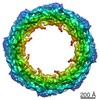
| Title | Human nuclear pore complex in HIV-1 infected T cells | |||||||||
 Map data Map data | Human Nuclear Pore Complex from HIV-1 infected T cells | |||||||||
 Sample Sample |
| |||||||||
| Biological species |  Homo sapiens (human) Homo sapiens (human) | |||||||||
| Method | subtomogram averaging / cryo EM / Resolution: 37.0 Å | |||||||||
 Authors Authors | Margiotta E / Beck M | |||||||||
| Funding support |  Germany, 1 items Germany, 1 items
| |||||||||
 Citation Citation | Journal: Cell / Year: 2021 Title: Cone-shaped HIV-1 capsids are transported through intact nuclear pores. Authors: Vojtech Zila / Erica Margiotta / Beata Turoňová / Thorsten G Müller / Christian E Zimmerli / Simone Mattei / Matteo Allegretti / Kathleen Börner / Jona Rada / Barbara Müller / Marina ...Authors: Vojtech Zila / Erica Margiotta / Beata Turoňová / Thorsten G Müller / Christian E Zimmerli / Simone Mattei / Matteo Allegretti / Kathleen Börner / Jona Rada / Barbara Müller / Marina Lusic / Hans-Georg Kräusslich / Martin Beck / Abstract: Human immunodeficiency virus (HIV-1) remains a major health threat. Viral capsid uncoating and nuclear import of the viral genome are critical for productive infection. The size of the HIV-1 capsid ...Human immunodeficiency virus (HIV-1) remains a major health threat. Viral capsid uncoating and nuclear import of the viral genome are critical for productive infection. The size of the HIV-1 capsid is generally believed to exceed the diameter of the nuclear pore complex (NPC), indicating that capsid uncoating has to occur prior to nuclear import. Here, we combined correlative light and electron microscopy with subtomogram averaging to capture the structural status of reverse transcription-competent HIV-1 complexes in infected T cells. We demonstrated that the diameter of the NPC in cellulo is sufficient for the import of apparently intact, cone-shaped capsids. Subsequent to nuclear import, we detected disrupted and empty capsid fragments, indicating that uncoating of the replication complex occurs by breaking the capsid open, and not by disassembly into individual subunits. Our data directly visualize a key step in HIV-1 replication and enhance our mechanistic understanding of the viral life cycle. #1: Journal: To Be PublishedTitle: Cone-shaped HIV-1 capsids are transported through intact nuclear pores Authors: Zila V / Margiotta E | |||||||||
| History |
|
- Structure visualization
| Movie |
 Movie viewer Movie viewer |
|---|---|
| Structure viewer | EM map: SurfViewMolmilJmol/JSmol |
| Supplemental images |

- Downloads & links
-EMDB archive
| Map data | emd_11967.map.gz | 2.5 MB | EMDB map data format | |
|---|---|---|---|---|
| Header (meta data) | emd-11967-v30.xmlemd-11967.xml | 9.4 KB 9.4 KB | Display Display | EMDB header |
| Images |  emd_11967.png emd_11967.png | 40.5 KB | ||
| Archive directory |  http://ftp.pdbj.org/pub/emdb/structures/EMD-11967ftp://ftp.pdbj.org/pub/emdb/structures/EMD-11967 http://ftp.pdbj.org/pub/emdb/structures/EMD-11967ftp://ftp.pdbj.org/pub/emdb/structures/EMD-11967 | HTTPS FTP |
-Related structure data
-Links
| EMDB pages | EMDB (EBI/PDBe) / EMDataResource |
|---|
-Map
| File | Download / File: emd_11967.map.gz / Format: CCP4 / Size: 11.4 MB / Type: IMAGE STORED AS FLOATING POINT NUMBER (4 BYTES) | ||||||||||||||||||||||||||||||||||||||||||||||||||||||||||||
|---|---|---|---|---|---|---|---|---|---|---|---|---|---|---|---|---|---|---|---|---|---|---|---|---|---|---|---|---|---|---|---|---|---|---|---|---|---|---|---|---|---|---|---|---|---|---|---|---|---|---|---|---|---|---|---|---|---|---|---|---|---|
| Annotation | Human Nuclear Pore Complex from HIV-1 infected T cells | ||||||||||||||||||||||||||||||||||||||||||||||||||||||||||||
| Projections & slices | Image control
Images are generated by Spider. | ||||||||||||||||||||||||||||||||||||||||||||||||||||||||||||
| Voxel size | X=Y=Z: 13.8 Å | ||||||||||||||||||||||||||||||||||||||||||||||||||||||||||||
| Density |
| ||||||||||||||||||||||||||||||||||||||||||||||||||||||||||||
| Symmetry | Space group: 1 | ||||||||||||||||||||||||||||||||||||||||||||||||||||||||||||
| Details | EMDB XML:
CCP4 map header:
| ||||||||||||||||||||||||||||||||||||||||||||||||||||||||||||
 Z (Sec.)
 Y (Row.)
 X (Col.)


-Supplemental data
- Sample components
-Entire : Nuclear pore complex from HIV-1 infected T cells
| Entire | Name: Nuclear pore complex from HIV-1 infected T cells |
|---|---|
| Components |
|
-Supramolecule #1: Nuclear pore complex from HIV-1 infected T cells
| Supramolecule | Name: Nuclear pore complex from HIV-1 infected T cells / type: complex / ID: 1 / Parent: 0 |
|---|---|
| Source (natural) | Organism: Homo sapiens (human) |
-Experimental details
-Structure determination
| Method | cryo EM |
|---|---|
 Processing Processing | subtomogram averaging |
| Aggregation state | cell |
-Sample preparation
| Buffer | pH: 7.4 / Details: RPMI 1640 medium with GlutaMAX |
|---|---|
| Grid | Model: Quantifoil / Material: GOLD / Mesh: 200 / Pretreatment - Type: GLOW DISCHARGE |
| Vitrification | Cryogen name: ETHANE / Chamber humidity: 100 % / Chamber temperature: 310.15 K / Instrument: LEICA EM GP |
- Electron microscopy
| Microscope | FEI TITAN KRIOS |
|---|---|
| Image recording | Film or detector model: GATAN K2 QUANTUM (4k x 4k) / Average electron dose: 4.0 e/Å2 |
| Electron beam | Acceleration voltage: 300 kV / Electron source:  FIELD EMISSION GUN FIELD EMISSION GUN |
| Electron optics | Illumination mode: OTHER / Imaging mode: OTHER / Nominal defocus max: 5.0 µm / Nominal defocus min: 2.0 µm / Nominal magnification: 42000 |
| Experimental equipment |  Model: Titan Krios / Image courtesy: FEI Company |
-Image processing
| Final reconstruction | Applied symmetry - Point group: C8 (8 fold cyclic) / Resolution.type: BY AUTHOR / Resolution: 37.0 Å / Resolution method: FSC 0.143 CUT-OFF / Number subtomograms used: 90 |
|---|---|
| Extraction | Number tomograms: 250 / Number images used: 99 |
| CTF correction | Details: novaCTF (Turonova et al., 2017) |
| Final angle assignment | Type: PROJECTION MATCHING |
