 Movie
Movie Controller

[English] 日本語
 Yorodumi
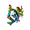
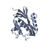
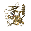
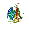
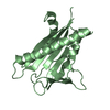
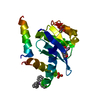
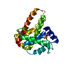
- PDB-7sv5: Crystal structure of SpaA-SLH/G109A in complex with 4,6-Pyr-beta-... -
+ Open data

- Basic information
| Entry | Database: PDB / ID: 7sv5 | ||||||||||||
|---|---|---|---|---|---|---|---|---|---|---|---|---|---|
| Title | Crystal structure of SpaA-SLH/G109A in complex with 4,6-Pyr-beta-D-ManNAc-(1->4)-beta-D-GlcNAcOMe | ||||||||||||
 Components Components | Surface (S-) layer glycoprotein | ||||||||||||
 Keywords Keywords | SUGAR BINDING PROTEIN / S-layer / SLH domain / Secondary cell wall polymer | ||||||||||||
| Function / homology | S-layer homology domain / S-layer homology (SLH) domain profile. / Copper resistance protein CopC/internalin, immunoglobulin-like / Chem-D2Y / Surface (S-) layer glycoprotein Function and homology information Function and homology information | ||||||||||||
| Biological species |  Paenibacillus alvei (bacteria) Paenibacillus alvei (bacteria) | ||||||||||||
| Method |  X-RAY DIFFRACTION / MOLECULAR REPLACEMENT / Resolution: 1.72 Å X-RAY DIFFRACTION / MOLECULAR REPLACEMENT / Resolution: 1.72 Å | ||||||||||||
 Authors Authors | Legg, M.S.G. / Evans, S.V. | ||||||||||||
| Funding support |  Austria, 3items Austria, 3items
| ||||||||||||
 Citation Citation | Journal: J.Biol.Chem. / Year: 2022 Title: The S-layer homology domains of Paenibacillus alvei surface protein SpaA bind to cell wall polysaccharide through the terminal monosaccharide residue. Authors: Legg, M.S.G. / Hager-Mair, F.F. / Krauter, S. / Gagnon, S.M.L. / Lopez-Guzman, A. / Lim, C. / Blaukopf, M. / Kosma, P. / Schaffer, C. / Evans, S.V. | ||||||||||||
| History |
|
- Structure visualization
| Structure viewer | Molecule: MolmilJmol/JSmol |
|---|
- Downloads & links
-Download
| PDBx/mmCIF format | 7sv5.cif.gz | 86.7 KB | Display | PDBx/mmCIF format |
|---|---|---|---|---|
| PDB format | pdb7sv5.ent.gz | 62.9 KB | Display | PDB format |
| PDBx/mmJSON format | 7sv5.json.gz | Tree view | PDBx/mmJSON format | |
| Others |  Other downloads Other downloads |
-Validation report
| Arichive directory | https://data.pdbj.org/pub/pdb/validation_reports/sv/7sv5ftp://data.pdbj.org/pub/pdb/validation_reports/sv/7sv5 | HTTPS FTP |
|---|
-Related structure data
| Related structure data |  7sv3C 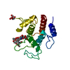 7sv4C  7sv6C 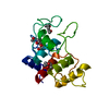 6cwnS C: citing same article ( S: Starting model for refinement |
|---|---|
| Similar structure data |


-Links
 PDBj
PDBj- Assembly
| Deposited unit | 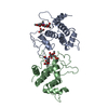
| ||||||||
|---|---|---|---|---|---|---|---|---|---|
| 1 | 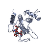
| ||||||||
| 2 | 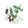
| ||||||||
| Unit cell |
| ||||||||
| Components on special symmetry positions |
|
-Components
| #1: Protein | Mass: 19798.266 Da / Num. of mol.: 2 / Fragment: SLH domains (UNP residues 21-193) / Mutation: G109A Source method: isolated from a genetically manipulated source Source: (gene. exp.) Paenibacillus alvei (bacteria) / Gene: spaA / Production host: #2: Chemical |   Mass: 92.094 Da / Num. of mol.: 2 / Source method: obtained synthetically / Formula: C3H8O3 Mass: 92.094 Da / Num. of mol.: 2 / Source method: obtained synthetically / Formula: C3H8O3#3: Chemical | 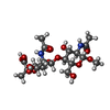  Mass: 508.474 Da / Num. of mol.: 2 / Source method: obtained synthetically / Formula: C20H32N2O13 / Feature type: SUBJECT OF INVESTIGATION Mass: 508.474 Da / Num. of mol.: 2 / Source method: obtained synthetically / Formula: C20H32N2O13 / Feature type: SUBJECT OF INVESTIGATION#4: Water | ChemComp-HOH / |  Mass: 18.015 Da / Num. of mol.: 303 / Source method: isolated from a natural source / Formula: H2O Mass: 18.015 Da / Num. of mol.: 303 / Source method: isolated from a natural source / Formula: H2OHas ligand of interest | Y | |
|---|
-Experimental details
-Experiment
| Experiment | Method: X-RAY DIFFRACTION / Number of used crystals: 1 |
|---|
- Sample preparation
| Crystal | Density Matthews: 2.16 Å3/Da / Density % sol: 43.09 % |
|---|---|
| Crystal grow | Temperature: 291.15 K / Method: vapor diffusion, sitting drop / pH: 7.5 / Details: 0.1 M HEPES pH 7.5, 25% (w/v) PEG 1000 |
-Data collection
| Diffraction | Mean temperature: 100 K / Serial crystal experiment: N | |||||||||||||||||||||||||||||||||||||||||||||||||||||||||||||||||||||||||||||||||||||||||||||||||||
|---|---|---|---|---|---|---|---|---|---|---|---|---|---|---|---|---|---|---|---|---|---|---|---|---|---|---|---|---|---|---|---|---|---|---|---|---|---|---|---|---|---|---|---|---|---|---|---|---|---|---|---|---|---|---|---|---|---|---|---|---|---|---|---|---|---|---|---|---|---|---|---|---|---|---|---|---|---|---|---|---|---|---|---|---|---|---|---|---|---|---|---|---|---|---|---|---|---|---|---|---|
| Diffraction source | Source: ROTATING ANODE / Type: RIGAKU MICROMAX-007 HF / Wavelength: 1.5418 Å | |||||||||||||||||||||||||||||||||||||||||||||||||||||||||||||||||||||||||||||||||||||||||||||||||||
| Detector | Type: DECTRIS PILATUS 200K / Detector: PIXEL / Date: Jun 1, 2019 | |||||||||||||||||||||||||||||||||||||||||||||||||||||||||||||||||||||||||||||||||||||||||||||||||||
| Radiation | Protocol: SINGLE WAVELENGTH / Monochromatic (M) / Laue (L): M / Scattering type: x-ray | |||||||||||||||||||||||||||||||||||||||||||||||||||||||||||||||||||||||||||||||||||||||||||||||||||
| Radiation wavelength | Wavelength: 1.5418 Å / Relative weight: 1 | |||||||||||||||||||||||||||||||||||||||||||||||||||||||||||||||||||||||||||||||||||||||||||||||||||
| Reflection | Resolution: 1.72→20 Å / Num. obs: 32683 / % possible obs: 90.3 % / Redundancy: 6.7 % / Rmerge(I) obs: 0.071 / Rpim(I) all: 0.029 / Rrim(I) all: 0.077 / Χ2: 0.677 / Net I/σ(I): 12.4 / Num. measured all: 217438 | |||||||||||||||||||||||||||||||||||||||||||||||||||||||||||||||||||||||||||||||||||||||||||||||||||
| Reflection shell | Diffraction-ID: 1
|
- Processing
| Software |
| ||||||||||||||||||||||||||||||||||||||||||||||||||||||||||||
|---|---|---|---|---|---|---|---|---|---|---|---|---|---|---|---|---|---|---|---|---|---|---|---|---|---|---|---|---|---|---|---|---|---|---|---|---|---|---|---|---|---|---|---|---|---|---|---|---|---|---|---|---|---|---|---|---|---|---|---|---|---|
| Refinement | Method to determine structure: MOLECULAR REPLACEMENT Starting model: 6CWN Resolution: 1.72→19.86 Å / Cor.coef. Fo:Fc: 0.964 / Cor.coef. Fo:Fc free: 0.961 / SU B: 2.531 / SU ML: 0.079 / Cross valid method: THROUGHOUT / σ(F): 0 / ESU R: 0.125 / ESU R Free: 0.108 / Stereochemistry target values: MAXIMUM LIKELIHOOD Details: HYDROGENS HAVE BEEN ADDED IN THE RIDING POSITIONS U VALUES : REFINED INDIVIDUALLY
| ||||||||||||||||||||||||||||||||||||||||||||||||||||||||||||
| Solvent computation | Ion probe radii: 0.8 Å / Shrinkage radii: 0.8 Å / VDW probe radii: 1.2 Å / Solvent model: MASK | ||||||||||||||||||||||||||||||||||||||||||||||||||||||||||||
| Displacement parameters | Biso max: 53.94 Å2 / Biso mean: 20.215 Å2 / Biso min: 12.39 Å2
| ||||||||||||||||||||||||||||||||||||||||||||||||||||||||||||
| Refinement step | Cycle: final / Resolution: 1.72→19.86 Å
| ||||||||||||||||||||||||||||||||||||||||||||||||||||||||||||
| Refine LS restraints |
| ||||||||||||||||||||||||||||||||||||||||||||||||||||||||||||
| LS refinement shell | Resolution: 1.72→1.765 Å / Rfactor Rfree error: 0 / Total num. of bins used: 20
|
