 Movie
Movie Controller

[English] 日本語
 Yorodumi
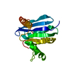
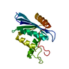
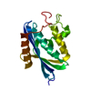
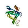
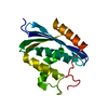
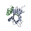
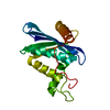
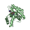
- PDB-6cwn: Crystal structure of SpaA-SLH/G109A in complex with 4,6-Pyr-beta-... -
+ Open data

- Basic information
| Entry | Database: PDB / ID: 6cwn | ||||||||||||
|---|---|---|---|---|---|---|---|---|---|---|---|---|---|
| Title | Crystal structure of SpaA-SLH/G109A in complex with 4,6-Pyr-beta-D-ManNAcOMe | ||||||||||||
 Components Components | Surface (S-) layer glycoprotein | ||||||||||||
 Keywords Keywords | SUGAR BINDING PROTEIN / Surface layer homology domain / Secondary cell wall polymer / S-layer / SLH / SCWP | ||||||||||||
| Function / homology | S-layer homology domain / S-layer homology (SLH) domain profile. / Copper resistance protein CopC/internalin, immunoglobulin-like / Chem-6LA / Surface (S-) layer glycoprotein Function and homology information Function and homology information | ||||||||||||
| Biological species |  Paenibacillus alvei (bacteria) Paenibacillus alvei (bacteria) | ||||||||||||
| Method |  X-RAY DIFFRACTION / SYNCHROTRON / MOLECULAR REPLACEMENT / Resolution: 1.53 Å X-RAY DIFFRACTION / SYNCHROTRON / MOLECULAR REPLACEMENT / Resolution: 1.53 Å | ||||||||||||
 Authors Authors | Blackler, R.J. / Evans, S.V. | ||||||||||||
| Funding support |  Canada, Canada,  Austria, 3items Austria, 3items
| ||||||||||||
 Citation Citation | Journal: Nat Commun / Year: 2018 Title: Structural basis of cell wall anchoring by SLH domains in Paenibacillus alvei. Authors: Blackler, R.J. / Lopez-Guzman, A. / Hager, F.F. / Janesch, B. / Martinz, G. / Gagnon, S.M.L. / Haji-Ghassemi, O. / Kosma, P. / Messner, P. / Schaffer, C. / Evans, S.V. | ||||||||||||
| History |
|
- Structure visualization
| Structure viewer | Molecule: MolmilJmol/JSmol |
|---|
- Downloads & links
-Download
| PDBx/mmCIF format | 6cwn.cif.gz | 88.7 KB | Display | PDBx/mmCIF format |
|---|---|---|---|---|
| PDB format | pdb6cwn.ent.gz | 65.5 KB | Display | PDB format |
| PDBx/mmJSON format | 6cwn.json.gz | Tree view | PDBx/mmJSON format | |
| Others |  Other downloads Other downloads |
-Validation report
| Arichive directory | https://data.pdbj.org/pub/pdb/validation_reports/cw/6cwnftp://data.pdbj.org/pub/pdb/validation_reports/cw/6cwn | HTTPS FTP |
|---|
-Related structure data
| Related structure data | 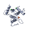 6cwcC  6cwfC 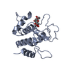 6cwhC 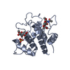 6cwiC  6cwlC  6cwmC 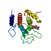 6cwrC C: citing same article ( |
|---|---|
| Similar structure data |

-Links
 PDBj
PDBj- Assembly
| Deposited unit | 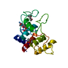
| ||||||||||||
|---|---|---|---|---|---|---|---|---|---|---|---|---|---|
| 1 |
| ||||||||||||
| Unit cell |
| ||||||||||||
| Components on special symmetry positions |
|
-Components
| #1: Protein | Mass: 19798.266 Da / Num. of mol.: 1 / Fragment: SLH domains (UNP residues 21-193) / Mutation: G109A Source method: isolated from a genetically manipulated source Source: (gene. exp.) Paenibacillus alvei (bacteria) / Gene: spaA / Plasmid: pET-22b / Production host: | ||
|---|---|---|---|
| #2: Sugar | ChemComp-6LA /   Type: D-saccharide / Mass: 305.281 Da / Num. of mol.: 1 / Source method: obtained synthetically / Formula: C12H19NO8 Type: D-saccharide / Mass: 305.281 Da / Num. of mol.: 1 / Source method: obtained synthetically / Formula: C12H19NO8 | ||
| #3: Chemical |   Mass: 118.174 Da / Num. of mol.: 2 / Source method: obtained synthetically / Formula: C6H14O2 / Comment: precipitant*YM Mass: 118.174 Da / Num. of mol.: 2 / Source method: obtained synthetically / Formula: C6H14O2 / Comment: precipitant*YM#4: Water | ChemComp-HOH / |  Mass: 18.015 Da / Num. of mol.: 176 / Source method: isolated from a natural source / Formula: H2O Mass: 18.015 Da / Num. of mol.: 176 / Source method: isolated from a natural source / Formula: H2O |
-Experimental details
-Experiment
| Experiment | Method: X-RAY DIFFRACTION / Number of used crystals: 1 |
|---|
- Sample preparation
| Crystal | Density Matthews: 2.36 Å3/Da / Density % sol: 47.94 % / Mosaicity: 0.95 ° |
|---|---|
| Crystal grow | Temperature: 289 K / Method: vapor diffusion, hanging drop Details: 0.1 M potassium thiocyanate, pH 6.8, 30% PEG2000 MME |
-Data collection
| Diffraction | Mean temperature: 100 K | |||||||||||||||||||||||||||||||||||||||||||||||||||||||||||||||||||||||||||||||||||||||||||||||||||||||||||||||||||||||||||||||||||||||||||||||||||||||||||||||||||||||||||||||||||||||||||||
|---|---|---|---|---|---|---|---|---|---|---|---|---|---|---|---|---|---|---|---|---|---|---|---|---|---|---|---|---|---|---|---|---|---|---|---|---|---|---|---|---|---|---|---|---|---|---|---|---|---|---|---|---|---|---|---|---|---|---|---|---|---|---|---|---|---|---|---|---|---|---|---|---|---|---|---|---|---|---|---|---|---|---|---|---|---|---|---|---|---|---|---|---|---|---|---|---|---|---|---|---|---|---|---|---|---|---|---|---|---|---|---|---|---|---|---|---|---|---|---|---|---|---|---|---|---|---|---|---|---|---|---|---|---|---|---|---|---|---|---|---|---|---|---|---|---|---|---|---|---|---|---|---|---|---|---|---|---|---|---|---|---|---|---|---|---|---|---|---|---|---|---|---|---|---|---|---|---|---|---|---|---|---|---|---|---|---|---|---|---|---|
| Diffraction source | Source: SYNCHROTRON / Site: CLSI / Beamline: 08ID-1 / Wavelength: 0.9795 Å | |||||||||||||||||||||||||||||||||||||||||||||||||||||||||||||||||||||||||||||||||||||||||||||||||||||||||||||||||||||||||||||||||||||||||||||||||||||||||||||||||||||||||||||||||||||||||||||
| Detector | Type: RAYONIX MX-300 / Detector: CCD / Date: Mar 31, 2016 | |||||||||||||||||||||||||||||||||||||||||||||||||||||||||||||||||||||||||||||||||||||||||||||||||||||||||||||||||||||||||||||||||||||||||||||||||||||||||||||||||||||||||||||||||||||||||||||
| Radiation | Monochromator: double crystal Si(111) / Protocol: SINGLE WAVELENGTH / Monochromatic (M) / Laue (L): M / Scattering type: x-ray | |||||||||||||||||||||||||||||||||||||||||||||||||||||||||||||||||||||||||||||||||||||||||||||||||||||||||||||||||||||||||||||||||||||||||||||||||||||||||||||||||||||||||||||||||||||||||||||
| Radiation wavelength | Wavelength: 0.9795 Å / Relative weight: 1 | |||||||||||||||||||||||||||||||||||||||||||||||||||||||||||||||||||||||||||||||||||||||||||||||||||||||||||||||||||||||||||||||||||||||||||||||||||||||||||||||||||||||||||||||||||||||||||||
| Reflection | Resolution: 1.53→50 Å / Num. obs: 27852 / % possible obs: 98.6 % / Redundancy: 4.3 % / Rmerge(I) obs: 0.044 / Rpim(I) all: 0.021 / Rrim(I) all: 0.049 / Χ2: 0.955 / Net I/av σ(I): 28.562 / Net I/σ(I): 23.5 / Num. measured all: 119834 | |||||||||||||||||||||||||||||||||||||||||||||||||||||||||||||||||||||||||||||||||||||||||||||||||||||||||||||||||||||||||||||||||||||||||||||||||||||||||||||||||||||||||||||||||||||||||||||
| Reflection shell | Diffraction-ID: 1 / Rejects: _
|
- Processing
| Software |
| ||||||||||||||||||||||||||||||||||||||||||||||||||||||||||||||||||||||||||||||||||||||||||
|---|---|---|---|---|---|---|---|---|---|---|---|---|---|---|---|---|---|---|---|---|---|---|---|---|---|---|---|---|---|---|---|---|---|---|---|---|---|---|---|---|---|---|---|---|---|---|---|---|---|---|---|---|---|---|---|---|---|---|---|---|---|---|---|---|---|---|---|---|---|---|---|---|---|---|---|---|---|---|---|---|---|---|---|---|---|---|---|---|---|---|---|
| Refinement | Method to determine structure: MOLECULAR REPLACEMENT / Resolution: 1.53→50 Å / Cor.coef. Fo:Fc: 0.977 / Cor.coef. Fo:Fc free: 0.966 / SU B: 1.782 / SU ML: 0.03 / Cross valid method: THROUGHOUT / σ(F): 0 / ESU R: 0.059 / ESU R Free: 0.056 Details: HYDROGENS HAVE BEEN ADDED IN THE RIDING POSITIONS U VALUES : WITH TLS ADDED
| ||||||||||||||||||||||||||||||||||||||||||||||||||||||||||||||||||||||||||||||||||||||||||
| Solvent computation | Ion probe radii: 0.8 Å / Shrinkage radii: 0.8 Å / VDW probe radii: 1.2 Å | ||||||||||||||||||||||||||||||||||||||||||||||||||||||||||||||||||||||||||||||||||||||||||
| Displacement parameters | Biso max: 92.91 Å2 / Biso mean: 17.079 Å2 / Biso min: 4.91 Å2
| ||||||||||||||||||||||||||||||||||||||||||||||||||||||||||||||||||||||||||||||||||||||||||
| Refinement step | Cycle: final / Resolution: 1.53→50 Å
| ||||||||||||||||||||||||||||||||||||||||||||||||||||||||||||||||||||||||||||||||||||||||||
| Refine LS restraints |
| ||||||||||||||||||||||||||||||||||||||||||||||||||||||||||||||||||||||||||||||||||||||||||
| LS refinement shell | Resolution: 1.532→1.572 Å / Total num. of bins used: 20
| ||||||||||||||||||||||||||||||||||||||||||||||||||||||||||||||||||||||||||||||||||||||||||
| Refinement TLS params. | Method: refined / Origin x: -10.1228 Å / Origin y: -1.9418 Å / Origin z: 10.3709 Å
|
