 Movie
Movie Controller

[English] 日本語
 Yorodumi
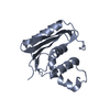
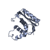
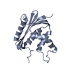
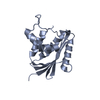
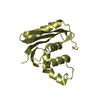
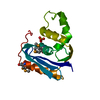
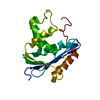
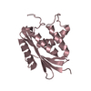
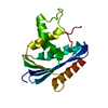
- PDB-1wsg: Co-crystal structure of E.coli RNase HI active site mutant (E48A/... -
+ Open data

- Basic information
| Entry | Database: PDB / ID: 1wsg | ||||||
|---|---|---|---|---|---|---|---|
| Title | Co-crystal structure of E.coli RNase HI active site mutant (E48A/D134N*) with Mn2+ | ||||||
 Components Components | Ribonuclease HI | ||||||
 Keywords Keywords | HYDROLASE / RNase H / active-site mutant / co-crystal structure with Mn2+ / metal fluctuation model | ||||||
| Function / homology |  Function and homology information Function and homology informationDNA replication, removal of RNA primer / ribonuclease H / RNA-DNA hybrid ribonuclease activity / endonuclease activity / nucleic acid binding / magnesium ion binding / cytoplasm Similarity search - Function | ||||||
| Biological species |  | ||||||
| Method |  X-RAY DIFFRACTION / SYNCHROTRON / MOLECULAR REPLACEMENT / Resolution: 2.2 Å X-RAY DIFFRACTION / SYNCHROTRON / MOLECULAR REPLACEMENT / Resolution: 2.2 Å | ||||||
 Authors Authors | Tsunaka, Y. / Takano, K. / Matsumura, H. / Yamagata, Y. / Kanaya, S. | ||||||
 Citation Citation | Journal: J.Mol.Biol. / Year: 2005 Title: Identification of Single Mn(2+) Binding Sites Required for Activation of the Mutant Proteins of E.coli RNase HI at Glu48 and/or Asp134 by X-ray Crystallography Authors: Tsunaka, Y. / Takano, K. / Matsumura, H. / Yamagata, Y. / Kanaya, S. | ||||||
| History |
|
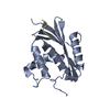
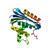
- Structure visualization
| Structure viewer | Molecule: MolmilJmol/JSmol |
|---|
- Downloads & links
-Download
| PDBx/mmCIF format | 1wsg.cif.gz | 137.2 KB | Display | PDBx/mmCIF format |
|---|---|---|---|---|
| PDB format | pdb1wsg.ent.gz | 108.7 KB | Display | PDB format |
| PDBx/mmJSON format | 1wsg.json.gz | Tree view | PDBx/mmJSON format | |
| Others |  Other downloads Other downloads |
-Validation report
| Arichive directory | https://data.pdbj.org/pub/pdb/validation_reports/ws/1wsgftp://data.pdbj.org/pub/pdb/validation_reports/ws/1wsg | HTTPS FTP |
|---|
-Related structure data


-Links
 PDBj
PDBj
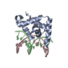
- Assembly
| Deposited unit | 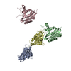
| ||||||||
|---|---|---|---|---|---|---|---|---|---|
| 1 | 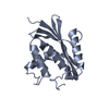
| ||||||||
| 2 | 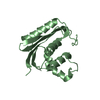
| ||||||||
| 3 | 
| ||||||||
| 4 | 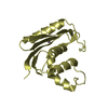
| ||||||||
| Unit cell |
|
-Components
| #1: Protein | Mass: 17505.873 Da / Num. of mol.: 4 / Mutation: E48A/K87A/D134N Source method: isolated from a genetically manipulated source Source: (gene. exp.) #2: Chemical |   Mass: 54.938 Da / Num. of mol.: 2 / Source method: obtained synthetically / Formula: Mn Mass: 54.938 Da / Num. of mol.: 2 / Source method: obtained synthetically / Formula: Mn#3: Water | ChemComp-HOH / |  Mass: 18.015 Da / Num. of mol.: 357 / Source method: isolated from a natural source / Formula: H2O Mass: 18.015 Da / Num. of mol.: 357 / Source method: isolated from a natural source / Formula: H2O |
|---|
-Experimental details
-Experiment
| Experiment | Method: X-RAY DIFFRACTION / Number of used crystals: 1 |
|---|
- Sample preparation
| Crystal | Density Matthews: 2.2 Å3/Da / Density % sol: 44.12 % |
|---|
-Data collection
| Diffraction source | Source: SYNCHROTRON / Site: SPring-8  / Beamline: BL38B1 / Wavelength: 1 Å / Beamline: BL38B1 / Wavelength: 1 Å |
|---|---|
| Detector | Type: ADSC QUANTUM 4 / Detector: CCD / Date: Sep 26, 2003 |
| Radiation | Protocol: SINGLE WAVELENGTH / Monochromatic (M) / Laue (L): M / Scattering type: x-ray |
| Radiation wavelength | Wavelength: 1 Å / Relative weight: 1 |
| Reflection | Resolution: 2.2→50 Å / Num. all: 32122 / Num. obs: 31685 / % possible obs: 98.6 % |
| Reflection shell | Highest resolution: 2.2 Å |
- Processing
| Software | Name: CNS / Classification: refinement | |||||||||||||||
|---|---|---|---|---|---|---|---|---|---|---|---|---|---|---|---|---|
| Refinement | Method to determine structure: MOLECULAR REPLACEMENT / Resolution: 2.2→50 Å
| |||||||||||||||
| Refinement step | Cycle: LAST / Resolution: 2.2→50 Å
|
