 Movie
Movie Controller

[English] 日本語
 Yorodumi
- PDB-7sq2: Reprocessed and refined structure of Phospholipase C-beta and Gq ... -
+ Open data

- Basic information
| Entry | Database: PDB / ID: 7sq2 | ||||||||||||||||||
|---|---|---|---|---|---|---|---|---|---|---|---|---|---|---|---|---|---|---|---|
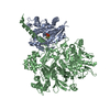
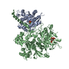
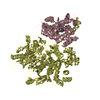
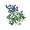
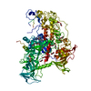
| Title | Reprocessed and refined structure of Phospholipase C-beta and Gq signaling complex | ||||||||||||||||||
 Components Components |
| ||||||||||||||||||
 Keywords Keywords | SIGNALING PROTEIN / G-protein signaling Phopholipase-C Hydrolase Guanine nucleotide-binding protein G(q) alpha | ||||||||||||||||||
| Function / homology |  Function and homology information Function and homology informationFatty Acids bound to GPR40 (FFAR1) regulate insulin secretion / PLC beta mediated events / Acetylcholine regulates insulin secretion / forebrain neuron development / regulation of melanocyte differentiation / Cooperation of PDCL (PhLP1) and TRiC/CCT in G-protein beta folding / Thromboxane signalling through TP receptor / phosphatidylinositol phospholipase C activity / Thrombin signalling through proteinase activated receptors (PARs) / adenylate cyclase-activating G protein-coupled cAMP receptor signaling pathway ...Fatty Acids bound to GPR40 (FFAR1) regulate insulin secretion / PLC beta mediated events / Acetylcholine regulates insulin secretion / forebrain neuron development / regulation of melanocyte differentiation / Cooperation of PDCL (PhLP1) and TRiC/CCT in G-protein beta folding / Thromboxane signalling through TP receptor / phosphatidylinositol phospholipase C activity / Thrombin signalling through proteinase activated receptors (PARs) / adenylate cyclase-activating G protein-coupled cAMP receptor signaling pathway / phosphoinositide phospholipase C / Turbulent (oscillatory, disturbed) flow shear stress activates signaling by PIEZO1 and integrins in endothelial cells / G-protein activation / G alpha (q) signalling events / phospholipase C-activating G protein-coupled acetylcholine receptor signaling pathway / endothelin receptor signaling pathway / Fatty Acids bound to GPR40 (FFAR1) regulate insulin secretion / Acetylcholine regulates insulin secretion / developmental pigmentation / phospholipase C-activating G protein-coupled glutamate receptor signaling pathway / phospholipase C-activating dopamine receptor signaling pathway / phosphatidylinositol metabolic process / regulation of systemic arterial blood pressure / PLC beta mediated events / ADP signalling through P2Y purinoceptor 1 / High laminar flow shear stress activates signaling by PIEZO1 and PECAM1:CDH5:KDR in endothelial cells / phospholipase C-activating serotonin receptor signaling pathway / phosphatidylinositol-4,5-bisphosphate phospholipase C activity / regulation of platelet activation / cranial skeletal system development / C-type glycerophospholipase activity / maternal behavior / regulation of canonical Wnt signaling pathway / embryonic digit morphogenesis / glutamate receptor signaling pathway / neuron remodeling / ligand-gated ion channel signaling pathway / alkylglycerophosphoethanolamine phosphodiesterase activity / Synthesis of IP3 and IP4 in the cytosol / phosphatidylinositol-mediated signaling / negative regulation of potassium ion transport / action potential / postsynaptic cytosol / lipid catabolic process / cellular response to acidic pH / enzyme regulator activity / release of sequestered calcium ion into cytosol / hormone-mediated signaling pathway / molecular function activator activity / GTPase activator activity / post-embryonic development / skeletal system development / neuropeptide signaling pathway / response to prostaglandin E / caveola / G protein-coupled receptor binding / regulation of blood pressure / G-protein beta/gamma-subunit complex binding / positive regulation of insulin secretion / G beta:gamma signalling through PLC beta / Presynaptic function of Kainate receptors / heterotrimeric G-protein complex / heart development / adenylate cyclase-activating G protein-coupled receptor signaling pathway / presynapse / cell body / G protein activity / Ca2+ pathway / nuclear membrane / molecular adaptor activity / phospholipase C-activating G protein-coupled receptor signaling pathway / G alpha (q) signalling events / Hydrolases; Acting on acid anhydrides; Acting on GTP to facilitate cellular and subcellular movement / calmodulin binding / protein stabilization / cadherin binding / G protein-coupled receptor signaling pathway / GTPase activity / calcium ion binding / dendrite / negative regulation of apoptotic process / GTP binding / protein-containing complex binding / Golgi apparatus / protein-containing complex / membrane / metal ion binding / nucleus / plasma membrane / cytoplasm / cytosol Similarity search - Function | ||||||||||||||||||
| Biological species |   Homo sapiens (human) Homo sapiens (human) | ||||||||||||||||||
| Method |  X-RAY DIFFRACTION / SYNCHROTRON / MOLECULAR REPLACEMENT / Resolution: 2.6 Å X-RAY DIFFRACTION / SYNCHROTRON / MOLECULAR REPLACEMENT / Resolution: 2.6 Å | ||||||||||||||||||
 Authors Authors | Endo-Streeter, S.T. / Sondek, J. / Harden, T.K. | ||||||||||||||||||
| Funding support |  United States, 5items United States, 5items
| ||||||||||||||||||
 Citation Citation | Journal: Science / Year: 2010 Title: Kinetic Scaffolding Mediated by a Phospholipase C-{beta} and Gq Signaling Complex Authors: Endo-Streeter, S.T. / Sondek, J. / Harden, T.K. | ||||||||||||||||||
| History |
|
- Structure visualization
| Structure viewer | Molecule: MolmilJmol/JSmol |
|---|
- Downloads & links
-Download
| PDBx/mmCIF format | 7sq2.cif.gz | 467.4 KB | Display | PDBx/mmCIF format |
|---|---|---|---|---|
| PDB format | pdb7sq2.ent.gz | 383 KB | Display | PDB format |
| PDBx/mmJSON format | 7sq2.json.gz | Tree view | PDBx/mmJSON format | |
| Others |  Other downloads Other downloads |
-Validation report
| Arichive directory | https://data.pdbj.org/pub/pdb/validation_reports/sq/7sq2ftp://data.pdbj.org/pub/pdb/validation_reports/sq/7sq2 | HTTPS FTP |
|---|
-Related structure data
| Related structure data |  3ohm S: Starting model for refinement |
|---|---|
| Similar structure data |

-Links
 PDBj
PDBj


- Assembly
| Deposited unit | 
| ||||||||
|---|---|---|---|---|---|---|---|---|---|
| 1 |
| ||||||||
| Unit cell |
|
-Components
-Protein , 2 types, 2 molecules AB
| #1: Protein | Mass: 38256.434 Da / Num. of mol.: 1 Source method: isolated from a genetically manipulated source Source: (gene. exp.)   Spodoptera frugiperda (fall armyworm) / References: UniProt: P21279 Spodoptera frugiperda (fall armyworm) / References: UniProt: P21279 |
|---|---|
| #2: Protein | Mass: 99865.281 Da / Num. of mol.: 1 Source method: isolated from a genetically manipulated source Source: (gene. exp.) Homo sapiens (human) / Gene: PLCB3 / Production host: Spodoptera frugiperda (fall armyworm)References: UniProt: Q01970, phosphoinositide phospholipase C |
-Non-polymers , 6 types, 227 molecules 


| #3: Chemical | ChemComp-ALF /  Mass: 102.975 Da / Num. of mol.: 1 / Source method: obtained synthetically / Formula: AlF4 / Feature type: SUBJECT OF INVESTIGATION Mass: 102.975 Da / Num. of mol.: 1 / Source method: obtained synthetically / Formula: AlF4 / Feature type: SUBJECT OF INVESTIGATION | ||||||||
|---|---|---|---|---|---|---|---|---|---|
| #4: Chemical |  Mass: 24.305 Da / Num. of mol.: 2 / Source method: obtained synthetically / Formula: Mg / Feature type: SUBJECT OF INVESTIGATION Mass: 24.305 Da / Num. of mol.: 2 / Source method: obtained synthetically / Formula: Mg / Feature type: SUBJECT OF INVESTIGATION#5: Chemical | ChemComp-GDP / |  Type: RNA linking / Mass: 443.201 Da / Num. of mol.: 1 / Source method: obtained synthetically / Formula: C10H15N5O11P2 / Feature type: SUBJECT OF INVESTIGATION / Comment: GDP, energy-carrying molecule*YM Type: RNA linking / Mass: 443.201 Da / Num. of mol.: 1 / Source method: obtained synthetically / Formula: C10H15N5O11P2 / Feature type: SUBJECT OF INVESTIGATION / Comment: GDP, energy-carrying molecule*YM#6: Chemical | ChemComp-ACT /  Mass: 59.044 Da / Num. of mol.: 7 / Source method: obtained synthetically / Formula: C2H3O2 Mass: 59.044 Da / Num. of mol.: 7 / Source method: obtained synthetically / Formula: C2H3O2#7: Chemical | ChemComp-CA / |  Mass: 40.078 Da / Num. of mol.: 1 / Source method: obtained synthetically / Formula: Ca / Feature type: SUBJECT OF INVESTIGATION Mass: 40.078 Da / Num. of mol.: 1 / Source method: obtained synthetically / Formula: Ca / Feature type: SUBJECT OF INVESTIGATION#8: Water | ChemComp-HOH / | Mass: 18.015 Da / Num. of mol.: 215 / Source method: isolated from a natural source / Formula: H2O |
-Details
| Has ligand of interest | Y |
|---|
-Experimental details
-Experiment
| Experiment | Method: X-RAY DIFFRACTION / Number of used crystals: 1 |
|---|
- Sample preparation
| Crystal | Density Matthews: 3.04 Å3/Da / Density % sol: 59.57 % |
|---|---|
| Crystal grow | Temperature: 291 K / Method: vapor diffusion, hanging drop / pH: 6 Details: 7% PEG 6000, 100 mM Hepes, 1 mM CaCl2, 100 mM magnesium acetate |
-Data collection
| Diffraction | Mean temperature: 100 K / Serial crystal experiment: N |
|---|---|
| Diffraction source | Source: SYNCHROTRON / Site: APS / Beamline: 22-ID / Wavelength: 1 Å |
| Detector | Type: MARMOSAIC 300 mm CCD / Detector: CCD / Date: Jun 8, 2010 / Details: Rosenbaum-Rock vertical focusing mirror |
| Radiation | Monochromator: Si 220. Rosenbaum-Rock double-crystal monochromator: liquid nitrogen cooled; sagittally focusing 2nd crystal, Rosenbaum-Rock vertical focusing mirror Protocol: SINGLE WAVELENGTH / Monochromatic (M) / Laue (L): M / Scattering type: x-ray |
| Radiation wavelength | Wavelength: 1 Å / Relative weight: 1 |
| Reflection | Resolution: 2.6→40.55 Å / Num. obs: 48454 / % possible obs: 99.49 % / Redundancy: 5.6 % / CC1/2: 0.905 / CC star: 0.975 / Rpim(I) all: 0.035 / Rrim(I) all: 0.084 / Χ2: 1.804 / Net I/σ(I): 32.5 |
| Reflection shell | Resolution: 2.6→2.666 Å / Redundancy: 5.5 % / Mean I/σ(I) obs: 2.67 / Num. unique obs: 2555 / CC1/2: 0.905 / CC star: 0.975 / Rpim(I) all: 0.304 / Rrim(I) all: 0.723 / Χ2: 0.957 / % possible all: 99.6 |
- Processing
| Software |
| ||||||||||||||||||||||||||||||||||||||||||||||||||||||||||||||||||||||||||||||||||||||||||||||||||||||||||||||||||||||||||||||||||||||||||||||||||||||||||||||||||||||||||||||||||||||
|---|---|---|---|---|---|---|---|---|---|---|---|---|---|---|---|---|---|---|---|---|---|---|---|---|---|---|---|---|---|---|---|---|---|---|---|---|---|---|---|---|---|---|---|---|---|---|---|---|---|---|---|---|---|---|---|---|---|---|---|---|---|---|---|---|---|---|---|---|---|---|---|---|---|---|---|---|---|---|---|---|---|---|---|---|---|---|---|---|---|---|---|---|---|---|---|---|---|---|---|---|---|---|---|---|---|---|---|---|---|---|---|---|---|---|---|---|---|---|---|---|---|---|---|---|---|---|---|---|---|---|---|---|---|---|---|---|---|---|---|---|---|---|---|---|---|---|---|---|---|---|---|---|---|---|---|---|---|---|---|---|---|---|---|---|---|---|---|---|---|---|---|---|---|---|---|---|---|---|---|---|---|---|---|
| Refinement | Method to determine structure: MOLECULAR REPLACEMENT Starting model: 3OHM 3ohm Resolution: 2.6→40.55 Å / Cor.coef. Fo:Fc: 0.959 / Cor.coef. Fo:Fc free: 0.925 / SU B: 29.23 / SU ML: 0.315 / Cross valid method: THROUGHOUT / ESU R: 0.444 / ESU R Free: 0.3 / Stereochemistry target values: MAXIMUM LIKELIHOOD / Details: HYDROGENS HAVE BEEN ADDED IN THE RIDING POSITIONS
| ||||||||||||||||||||||||||||||||||||||||||||||||||||||||||||||||||||||||||||||||||||||||||||||||||||||||||||||||||||||||||||||||||||||||||||||||||||||||||||||||||||||||||||||||||||||
| Solvent computation | Ion probe radii: 0.8 Å / Shrinkage radii: 0.8 Å / VDW probe radii: 1.2 Å / Solvent model: MASK | ||||||||||||||||||||||||||||||||||||||||||||||||||||||||||||||||||||||||||||||||||||||||||||||||||||||||||||||||||||||||||||||||||||||||||||||||||||||||||||||||||||||||||||||||||||||
| Displacement parameters | Biso mean: 91.472 Å2
| ||||||||||||||||||||||||||||||||||||||||||||||||||||||||||||||||||||||||||||||||||||||||||||||||||||||||||||||||||||||||||||||||||||||||||||||||||||||||||||||||||||||||||||||||||||||
| Refinement step | Cycle: 1 / Resolution: 2.6→40.55 Å
| ||||||||||||||||||||||||||||||||||||||||||||||||||||||||||||||||||||||||||||||||||||||||||||||||||||||||||||||||||||||||||||||||||||||||||||||||||||||||||||||||||||||||||||||||||||||
| Refine LS restraints |
|