 Movie
Movie Controller

[English] 日本語
 Yorodumi
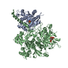
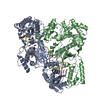
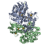
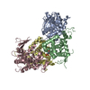
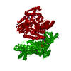
- PDB-4qj4: Structure of a fragment of human phospholipase C-beta3 delta472-5... -
+ Open data

- Basic information
| Entry | Database: PDB / ID: 4qj4 | ||||||
|---|---|---|---|---|---|---|---|
| Title | Structure of a fragment of human phospholipase C-beta3 delta472-569, bound to IP3 and in complex with Galphaq | ||||||
 Components Components |
| ||||||
 Keywords Keywords | SIGNALING PROTEIN/HYDROLASE / GTP-BINDING PROTEIN ALPHA SUBUNITS / PHOSPHOLIPASE C BETA / PH DOMAIN / EF HAND / C2 DOMAIN / TIM BARREL DOMAIN / GTP HYDROLYSIS / G-PROTEIN SIGNALING / LIPASE / CALCIUM BINDING / GTP BINDING / PHOSPHOLIPIDS / membrane / SIGNALING PROTEIN-HYDROLASE complex | ||||||
| Function / homology |  Function and homology information Function and homology informationFatty Acids bound to GPR40 (FFAR1) regulate insulin secretion / PLC beta mediated events / Acetylcholine regulates insulin secretion / forebrain neuron development / regulation of melanocyte differentiation / Cooperation of PDCL (PhLP1) and TRiC/CCT in G-protein beta folding / Thromboxane signalling through TP receptor / phosphatidylinositol phospholipase C activity / Thrombin signalling through proteinase activated receptors (PARs) / adenylate cyclase-activating G protein-coupled cAMP receptor signaling pathway ...Fatty Acids bound to GPR40 (FFAR1) regulate insulin secretion / PLC beta mediated events / Acetylcholine regulates insulin secretion / forebrain neuron development / regulation of melanocyte differentiation / Cooperation of PDCL (PhLP1) and TRiC/CCT in G-protein beta folding / Thromboxane signalling through TP receptor / phosphatidylinositol phospholipase C activity / Thrombin signalling through proteinase activated receptors (PARs) / adenylate cyclase-activating G protein-coupled cAMP receptor signaling pathway / regulation of systemic arterial blood pressure / phosphoinositide phospholipase C / Turbulent (oscillatory, disturbed) flow shear stress activates signaling by PIEZO1 and integrins in endothelial cells / G-protein activation / G alpha (q) signalling events / phospholipase C-activating G protein-coupled acetylcholine receptor signaling pathway / endothelin receptor signaling pathway / Fatty Acids bound to GPR40 (FFAR1) regulate insulin secretion / phospholipase C-activating dopamine receptor signaling pathway / Acetylcholine regulates insulin secretion / developmental pigmentation / phospholipase C-activating G protein-coupled glutamate receptor signaling pathway / phosphatidylinositol metabolic process / ADP signalling through P2Y purinoceptor 1 / High laminar flow shear stress activates signaling by PIEZO1 and PECAM1:CDH5:KDR in endothelial cells / phospholipase C-activating serotonin receptor signaling pathway / cranial skeletal system development / phosphatidylinositol-4,5-bisphosphate phospholipase C activity / regulation of platelet activation / PLC beta mediated events / C-type glycerophospholipase activity / maternal behavior / regulation of canonical Wnt signaling pathway / embryonic digit morphogenesis / glutamate receptor signaling pathway / neuron remodeling / ligand-gated ion channel signaling pathway / alkylglycerophosphoethanolamine phosphodiesterase activity / Synthesis of IP3 and IP4 in the cytosol / phosphatidylinositol-mediated signaling / negative regulation of potassium ion transport / action potential / postsynaptic cytosol / lipid catabolic process / hormone-mediated signaling pathway / cellular response to acidic pH / release of sequestered calcium ion into cytosol / enzyme regulator activity / post-embryonic development / molecular function activator activity / GTPase activator activity / skeletal system development / neuropeptide signaling pathway / response to prostaglandin E / caveola / G protein-coupled receptor binding / regulation of blood pressure / G-protein beta/gamma-subunit complex binding / positive regulation of insulin secretion / G beta:gamma signalling through PLC beta / Presynaptic function of Kainate receptors / heart development / heterotrimeric G-protein complex / presynapse / adenylate cyclase-activating G protein-coupled receptor signaling pathway / cell body / nuclear membrane / G protein activity / Ca2+ pathway / molecular adaptor activity / G alpha (q) signalling events / phospholipase C-activating G protein-coupled receptor signaling pathway / Hydrolases; Acting on acid anhydrides; Acting on GTP to facilitate cellular and subcellular movement / calmodulin binding / protein stabilization / cadherin binding / G protein-coupled receptor signaling pathway / GTPase activity / calcium ion binding / dendrite / negative regulation of apoptotic process / GTP binding / protein-containing complex binding / Golgi apparatus / protein-containing complex / membrane / metal ion binding / nucleus / plasma membrane / cytosol / cytoplasm Similarity search - Function | ||||||
| Biological species |   Homo sapiens (human) Homo sapiens (human) | ||||||
| Method |  X-RAY DIFFRACTION / SYNCHROTRON / MOLECULAR REPLACEMENT / Resolution: 3.3 Å X-RAY DIFFRACTION / SYNCHROTRON / MOLECULAR REPLACEMENT / Resolution: 3.3 Å | ||||||
 Authors Authors | Lyon, A.M. / Tesmer, J.J.G. | ||||||
 Citation Citation | Journal: Structure / Year: 2014 Title: Molecular mechanisms of phospholipase C beta 3 autoinhibition. Authors: Lyon, A.M. / Begley, J.A. / Manett, T.D. / Tesmer, J.J. | ||||||
| History |
|
- Structure visualization
| Structure viewer | Molecule: MolmilJmol/JSmol |
|---|
- Downloads & links
-Download
| PDBx/mmCIF format | 4qj4.cif.gz | 452.1 KB | Display | PDBx/mmCIF format |
|---|---|---|---|---|
| PDB format | pdb4qj4.ent.gz | 366.8 KB | Display | PDB format |
| PDBx/mmJSON format | 4qj4.json.gz | Tree view | PDBx/mmJSON format | |
| Others |  Other downloads Other downloads |
-Validation report
| Arichive directory | https://data.pdbj.org/pub/pdb/validation_reports/qj/4qj4ftp://data.pdbj.org/pub/pdb/validation_reports/qj/4qj4 | HTTPS FTP |
|---|
-Related structure data
| Related structure data | 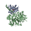 4qj3C  4qj5C  3ohm C: citing same article ( S: Starting model for refinement |
|---|---|
| Similar structure data |


-Links
 PDBj
PDBj


- Assembly
| Deposited unit | 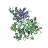
| ||||||||
|---|---|---|---|---|---|---|---|---|---|
| 1 |
| ||||||||
| Unit cell |
|
-Components
-Protein , 2 types, 2 molecules AB
| #1: Protein | Mass: 44711.590 Da / Num. of mol.: 1 / Fragment: UNP residues 7-359 Source method: isolated from a genetically manipulated source Source: (gene. exp.)  TRICHOPLUSIA NI (cabbage looper) / References: UniProt: P21279 TRICHOPLUSIA NI (cabbage looper) / References: UniProt: P21279 |
|---|---|
| #2: Protein | Mass: 90294.156 Da / Num. of mol.: 1 / Fragment: UNP residues 10-891 Source method: isolated from a genetically manipulated source Source: (gene. exp.) Homo sapiens (human) / Gene: PLCB3 / Plasmid: pFastBacDual / Production host: TRICHOPLUSIA NI (cabbage looper)References: UniProt: Q01970, phosphoinositide phospholipase C |
-Non-polymers , 6 types, 17 molecules 


| #3: Chemical | ChemComp-GDP /  Type: RNA linking / Mass: 443.201 Da / Num. of mol.: 1 / Source method: obtained synthetically / Formula: C10H15N5O11P2 / Comment: GDP, energy-carrying molecule*YM Type: RNA linking / Mass: 443.201 Da / Num. of mol.: 1 / Source method: obtained synthetically / Formula: C10H15N5O11P2 / Comment: GDP, energy-carrying molecule*YM |
|---|---|
| #4: Chemical | ChemComp-ALF /  Mass: 102.975 Da / Num. of mol.: 1 / Source method: obtained synthetically / Formula: AlF4 Mass: 102.975 Da / Num. of mol.: 1 / Source method: obtained synthetically / Formula: AlF4 |
| #5: Chemical | ChemComp-MG /  Mass: 24.305 Da / Num. of mol.: 1 / Source method: obtained synthetically / Formula: Mg Mass: 24.305 Da / Num. of mol.: 1 / Source method: obtained synthetically / Formula: Mg |
| #6: Chemical | ChemComp-CA /  Mass: 40.078 Da / Num. of mol.: 1 / Source method: obtained synthetically / Formula: Ca Mass: 40.078 Da / Num. of mol.: 1 / Source method: obtained synthetically / Formula: Ca |
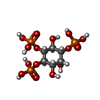
| #7: Chemical | ChemComp-I3P /  Mass: 420.096 Da / Num. of mol.: 1 / Source method: obtained synthetically / Formula: C6H15O15P3 Mass: 420.096 Da / Num. of mol.: 1 / Source method: obtained synthetically / Formula: C6H15O15P3 |
| #8: Water | ChemComp-HOH / Mass: 18.015 Da / Num. of mol.: 12 / Source method: isolated from a natural source / Formula: H2O |
-Experimental details
-Experiment
| Experiment | Method: X-RAY DIFFRACTION / Number of used crystals: 2 |
|---|
- Sample preparation
| Crystal | Density Matthews: 3.03 Å3/Da / Density % sol: 59.34 % |
|---|---|
| Crystal grow | Temperature: 277.15 K / Method: vapor diffusion, hanging drop / pH: 6.5 Details: 100 mM BisTris, 200 mM NaCl, 8% (v/v) PEG 3350, pH 6.5, VAPOR DIFFUSION, HANGING DROP, temperature 277.15K |
-Data collection
| Diffraction | Mean temperature: 110 K |
|---|---|
| Diffraction source | Source: SYNCHROTRON / Site: APS  / Beamline: 21-ID-D / Wavelength: 0.979 Å / Beamline: 21-ID-D / Wavelength: 0.979 Å |
| Detector | Type: MARMOSAIC 300 mm CCD / Detector: CCD / Date: Apr 18, 2014 |
| Radiation | Monochromator: KOHZU MONOCHROMATOR, CRYSTAL MONOCHROMATOR / Protocol: SINGLE WAVELENGTH / Monochromatic (M) / Laue (L): M / Scattering type: x-ray |
| Radiation wavelength | Wavelength: 0.979 Å / Relative weight: 1 |
| Reflection | Resolution: 3.3→30 Å / Num. all: 21343 / Num. obs: 21289 / % possible obs: 85.6 % / Observed criterion σ(F): -2000 / Observed criterion σ(I): -2 / Redundancy: 3.7 % / Rmerge(I) obs: 0.221 / Net I/σ(I): 8.71 |
| Reflection shell | Resolution: 3.3→3.36 Å / Redundancy: 2.7 % / Rmerge(I) obs: 0.48 / Mean I/σ(I) obs: 3.27 / % possible all: 64.6 |
- Processing
| Software |
| |||||||||||||||||||||||||||||||||||||||||||||||||||||||||||||||||||||||||||||||||||||||||||||||||||||||||
|---|---|---|---|---|---|---|---|---|---|---|---|---|---|---|---|---|---|---|---|---|---|---|---|---|---|---|---|---|---|---|---|---|---|---|---|---|---|---|---|---|---|---|---|---|---|---|---|---|---|---|---|---|---|---|---|---|---|---|---|---|---|---|---|---|---|---|---|---|---|---|---|---|---|---|---|---|---|---|---|---|---|---|---|---|---|---|---|---|---|---|---|---|---|---|---|---|---|---|---|---|---|---|---|---|---|---|
| Refinement | Method to determine structure: MOLECULAR REPLACEMENT Starting model: PDB ENTRY 3OHM 3ohm Resolution: 3.3→29.4 Å / Cor.coef. Fo:Fc: 0.91 / Cor.coef. Fo:Fc free: 0.851 / SU B: 62.713 / SU ML: 0.458 / Cross valid method: THROUGHOUT / σ(F): 0 / ESU R Free: 0.614 / Stereochemistry target values: MAXIMUM LIKELIHOOD / Details: HYDROGENS HAVE BEEN ADDED IN THE RIDING POSITIONS
| |||||||||||||||||||||||||||||||||||||||||||||||||||||||||||||||||||||||||||||||||||||||||||||||||||||||||
| Solvent computation | Ion probe radii: 0.8 Å / Shrinkage radii: 0.8 Å / VDW probe radii: 1.2 Å / Solvent model: MASK | |||||||||||||||||||||||||||||||||||||||||||||||||||||||||||||||||||||||||||||||||||||||||||||||||||||||||
| Displacement parameters | Biso mean: 72.003 Å2
| |||||||||||||||||||||||||||||||||||||||||||||||||||||||||||||||||||||||||||||||||||||||||||||||||||||||||
| Refinement step | Cycle: LAST / Resolution: 3.3→29.4 Å
| |||||||||||||||||||||||||||||||||||||||||||||||||||||||||||||||||||||||||||||||||||||||||||||||||||||||||
| Refine LS restraints |
| |||||||||||||||||||||||||||||||||||||||||||||||||||||||||||||||||||||||||||||||||||||||||||||||||||||||||
| LS refinement shell | Resolution: 3.3→3.361 Å / Total num. of bins used: 20
| |||||||||||||||||||||||||||||||||||||||||||||||||||||||||||||||||||||||||||||||||||||||||||||||||||||||||
| Refinement TLS params. | Method: refined / Refine-ID: X-RAY DIFFRACTION
| |||||||||||||||||||||||||||||||||||||||||||||||||||||||||||||||||||||||||||||||||||||||||||||||||||||||||
| Refinement TLS group |
|
