[English] 日本語
Yorodumi
- PDB-4gnk: Crystal structure of Galphaq in complex with full-length human PL... -

+
Open data


ID or keywords:

Loading...

-
Basic information

Entry
Database: PDB / ID: 4gnk
TitleCrystal structure of Galphaq in complex with full-length human PLCbeta3
Components
  • (1-phosphatidylinositol 4,5-bisphosphate phosphodiesterase beta- ...) x 2
  • Guanine nucleotide-binding protein G(q) subunit alpha
KeywordsGTP-BINDING PROTEIN/HYDROLASE / GTP-binding protein alpha subunits / phospholipase C beta / coiled-coil domain / PH DOMAIN / EF HAND / C2 DOMAIN / TIM BARREL DOMAIN / phospholipase / GTP hydrolysis / G-protein signaling / membrane targeting / lipase / hydrolase / calcium binding / GTP binding / phospholipids / GTP-BINDING PROTEIN-HYDROLASE complex
Function / homology
Function and homology information


Fatty Acids bound to GPR40 (FFAR1) regulate insulin secretion / Acetylcholine regulates insulin secretion / PLC beta mediated events / forebrain neuron development / regulation of melanocyte differentiation / Cooperation of PDCL (PhLP1) and TRiC/CCT in G-protein beta folding / Thromboxane signalling through TP receptor / Thrombin signalling through proteinase activated receptors (PARs) / G-protein activation / phosphoinositide phospholipase C ...Fatty Acids bound to GPR40 (FFAR1) regulate insulin secretion / Acetylcholine regulates insulin secretion / PLC beta mediated events / forebrain neuron development / regulation of melanocyte differentiation / Cooperation of PDCL (PhLP1) and TRiC/CCT in G-protein beta folding / Thromboxane signalling through TP receptor / Thrombin signalling through proteinase activated receptors (PARs) / G-protein activation / phosphoinositide phospholipase C / Fatty Acids bound to GPR40 (FFAR1) regulate insulin secretion / G alpha (q) signalling events / Acetylcholine regulates insulin secretion / endothelin receptor signaling pathway / ion channel modulating, G protein-coupled receptor signaling pathway / phospholipase C-activating G protein-coupled acetylcholine receptor signaling pathway / developmental pigmentation / phosphatidylinositol metabolic process / PLC beta mediated events / phospholipase C-activating dopamine receptor signaling pathway / cranial skeletal system development / ADP signalling through P2Y purinoceptor 1 / regulation of systemic arterial blood pressure / regulation of platelet activation / phosphatidylinositol phospholipase C activity / phospholipase C activity / maternal behavior / glutamate receptor signaling pathway / alkylglycerophosphoethanolamine phosphodiesterase activity / embryonic digit morphogenesis / postsynaptic cytosol / neuron remodeling / Synthesis of IP3 and IP4 in the cytosol / action potential / ligand-gated ion channel signaling pathway / negative regulation of potassium ion transport / phosphatidylinositol-mediated signaling / enzyme regulator activity / lipid catabolic process / release of sequestered calcium ion into cytosol / GTPase activator activity / positive regulation of smooth muscle cell proliferation / G protein activity / post-embryonic development / skeletal system development / molecular function activator activity / caveola / G protein-coupled receptor binding / regulation of blood pressure / G-protein beta/gamma-subunit complex binding / G beta:gamma signalling through PLC beta / Presynaptic function of Kainate receptors / positive regulation of insulin secretion / adenylate cyclase-activating G protein-coupled receptor signaling pathway / heterotrimeric G-protein complex / Ca2+ pathway / heart development / phospholipase C-activating G protein-coupled receptor signaling pathway / cell body / nuclear membrane / G alpha (q) signalling events / molecular adaptor activity / calmodulin binding / cadherin binding / G protein-coupled receptor signaling pathway / GTPase activity / synapse / dendrite / calcium ion binding / negative regulation of apoptotic process / protein-containing complex binding / GTP binding / Golgi apparatus / protein-containing complex / membrane / nucleus / metal ion binding / plasma membrane / cytoplasm / cytosol
Similarity search - Function
Phospholipase C Beta; Chain: A / Phospholipase C beta, distal C-terminal domain / PH-domain like - #240 / Phospholipase C-beta, C-terminal domain / PLC-beta C terminal / PLC-beta, PH domain / Phospholipase C-beta, C-terminal domain superfamily / PH domain / Phosphatidylinositol-4, 5-bisphosphate phosphodiesterase beta / Phosphoinositide-specific phospholipase C, EF-hand-like domain ...Phospholipase C Beta; Chain: A / Phospholipase C beta, distal C-terminal domain / PH-domain like - #240 / Phospholipase C-beta, C-terminal domain / PLC-beta C terminal / PLC-beta, PH domain / Phospholipase C-beta, C-terminal domain superfamily / PH domain / Phosphatidylinositol-4, 5-bisphosphate phosphodiesterase beta / Phosphoinositide-specific phospholipase C, EF-hand-like domain / Phosphoinositide-specific phospholipase C, efhand-like / G-protein alpha subunit, group Q / Phosphoinositide phospholipase C family / Phospholipase C, phosphatidylinositol-specific, Y domain / Phosphatidylinositol-specific phospholipase C, Y domain / Phosphatidylinositol-specific phospholipase Y-box domain profile. / Phospholipase C, catalytic domain (part); domain Y / Phosphatidylinositol (PI) phosphodiesterase / Phosphatidylinositol-specific phospholipase C, X domain / Phosphatidylinositol-specific phospholipase C, X domain / Phospholipase C, catalytic domain (part); domain X / Phosphatidylinositol-specific phospholipase X-box domain profile. / GI Alpha 1, domain 2-like / GI Alpha 1, domain 2-like / PLC-like phosphodiesterase, TIM beta/alpha-barrel domain superfamily / C2 domain / C2 domain / Protein kinase C conserved region 2 (CalB) / C2 domain / C2 domain profile. / PH-domain like / EF-hand / Recoverin; domain 1 / C2 domain superfamily / G-alpha domain profile. / Guanine nucleotide binding protein (G-protein), alpha subunit / G protein alpha subunit, helical insertion / G-protein alpha subunit / G protein alpha subunit / EF-hand domain pair / P-loop containing nucleotide triphosphate hydrolases / Roll / TIM Barrel / Alpha-Beta Barrel / Up-down Bundle / Immunoglobulin-like / Sandwich / Rossmann fold / P-loop containing nucleoside triphosphate hydrolase / Orthogonal Bundle / 3-Layer(aba) Sandwich / Mainly Beta / Mainly Alpha / Alpha Beta
Similarity search - Domain/homology
TETRAFLUOROALUMINATE ION / GUANOSINE-5'-DIPHOSPHATE / Guanine nucleotide-binding protein G(q) subunit alpha / 1-phosphatidylinositol 4,5-bisphosphate phosphodiesterase beta-3
Similarity search - Component
Biological speciesMus musculus (house mouse)
Homo sapiens (human)
MethodX-RAY DIFFRACTION / SYNCHROTRON / MOLECULAR REPLACEMENT / Resolution: 4 Å
AuthorsLyon, A.M. / Tesmer, J.J.G.
Citation
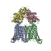
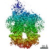
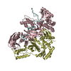
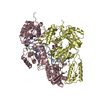
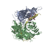
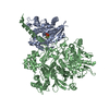
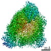
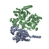
Journal: Nat Struct Mol Biol / Year: 2013
Title: Full-length Gα(q)-phospholipase C-β3 structure reveals interfaces of the C-terminal coiled-coil domain.
Authors: Angeline M Lyon / Somnath Dutta / Cassandra A Boguth / Georgios Skiniotis / John J G Tesmer /
Abstract: Phospholipase C-β (PLCβ) is directly activated by Gαq, but the molecular basis for how its distal C-terminal domain (CTD) contributes to maximal activity is poorly understood. Herein we present ...Phospholipase C-β (PLCβ) is directly activated by Gαq, but the molecular basis for how its distal C-terminal domain (CTD) contributes to maximal activity is poorly understood. Herein we present both the crystal structure and cryo-EM three-dimensional reconstructions of human full-length PLCβ3 in complex with mouse Gαq. The distal CTD forms an extended monomeric helical bundle consisting of three antiparallel segments with structural similarity to membrane-binding bin-amphiphysin-Rvs (BAR) domains. Sequence conservation of the distal CTD suggests putative membrane and protein interaction sites, the latter of which bind the N-terminal helix of Gαq in both the crystal structure and cryo-EM reconstructions. Functional analysis suggests that the distal CTD has roles in membrane targeting and in optimizing the orientation of the catalytic core at the membrane for maximal rates of lipid hydrolysis.
History
DepositionAug 17, 2012Deposition site: RCSB / Processing site: RCSB
Revision 1.0Feb 6, 2013Provider: repository / Type: Initial release
Revision 1.1Sep 4, 2013Group: Database references
Revision 1.2Sep 13, 2023Group: Data collection / Database references ...Data collection / Database references / Derived calculations / Refinement description
Category: chem_comp_atom / chem_comp_bond ...chem_comp_atom / chem_comp_bond / database_2 / pdbx_initial_refinement_model / pdbx_struct_conn_angle / struct_conn / struct_ref_seq_dif / struct_site
Item: _database_2.pdbx_DOI / _database_2.pdbx_database_accession ..._database_2.pdbx_DOI / _database_2.pdbx_database_accession / _pdbx_struct_conn_angle.ptnr1_auth_asym_id / _pdbx_struct_conn_angle.ptnr1_auth_comp_id / _pdbx_struct_conn_angle.ptnr1_auth_seq_id / _pdbx_struct_conn_angle.ptnr1_label_asym_id / _pdbx_struct_conn_angle.ptnr1_label_atom_id / _pdbx_struct_conn_angle.ptnr1_label_comp_id / _pdbx_struct_conn_angle.ptnr1_label_seq_id / _pdbx_struct_conn_angle.ptnr2_auth_asym_id / _pdbx_struct_conn_angle.ptnr2_auth_comp_id / _pdbx_struct_conn_angle.ptnr2_auth_seq_id / _pdbx_struct_conn_angle.ptnr2_label_asym_id / _pdbx_struct_conn_angle.ptnr2_label_atom_id / _pdbx_struct_conn_angle.ptnr2_label_comp_id / _pdbx_struct_conn_angle.ptnr3_auth_asym_id / _pdbx_struct_conn_angle.ptnr3_auth_comp_id / _pdbx_struct_conn_angle.ptnr3_auth_seq_id / _pdbx_struct_conn_angle.ptnr3_label_asym_id / _pdbx_struct_conn_angle.ptnr3_label_atom_id / _pdbx_struct_conn_angle.ptnr3_label_comp_id / _pdbx_struct_conn_angle.ptnr3_label_seq_id / _pdbx_struct_conn_angle.value / _struct_conn.pdbx_dist_value / _struct_conn.ptnr1_auth_asym_id / _struct_conn.ptnr1_auth_comp_id / _struct_conn.ptnr1_auth_seq_id / _struct_conn.ptnr1_label_asym_id / _struct_conn.ptnr1_label_atom_id / _struct_conn.ptnr1_label_comp_id / _struct_conn.ptnr1_label_seq_id / _struct_conn.ptnr2_auth_asym_id / _struct_conn.ptnr2_auth_comp_id / _struct_conn.ptnr2_auth_seq_id / _struct_conn.ptnr2_label_asym_id / _struct_conn.ptnr2_label_atom_id / _struct_conn.ptnr2_label_comp_id / _struct_ref_seq_dif.details / _struct_site.pdbx_auth_asym_id / _struct_site.pdbx_auth_comp_id / _struct_site.pdbx_auth_seq_id

-
Structure visualization

Structure viewerMolecule:
MolmilJmol/JSmol

Downloads & links

-
Assembly

Deposited unit
A: Guanine nucleotide-binding protein G(q) subunit alpha
B: 1-phosphatidylinositol 4,5-bisphosphate phosphodiesterase beta-3
C: Guanine nucleotide-binding protein G(q) subunit alpha
D: 1-phosphatidylinositol 4,5-bisphosphate phosphodiesterase beta-3
E: 1-phosphatidylinositol 4,5-bisphosphate phosphodiesterase beta-3
hetero molecules


Theoretical massNumber of molelcules
Total (without water)393,28113
Polymers392,0605
Non-polymers1,2218
Water1086
1
A: Guanine nucleotide-binding protein G(q) subunit alpha
B: 1-phosphatidylinositol 4,5-bisphosphate phosphodiesterase beta-3
hetero molecules


Theoretical massNumber of molelcules
Total (without water)181,4676
Polymers180,8562
Non-polymers6114
Water181
TypeNameSymmetry operationNumber
identity operation1_555x,y,z1
Buried area4570 Å2
ΔGint-40 kcal/mol
Surface area49960 Å2
MethodPISA
2
C: Guanine nucleotide-binding protein G(q) subunit alpha
D: 1-phosphatidylinositol 4,5-bisphosphate phosphodiesterase beta-3
hetero molecules


Theoretical massNumber of molelcules
Total (without water)181,4676
Polymers180,8562
Non-polymers6114
Water181
TypeNameSymmetry operationNumber
identity operation1_555x,y,z1
Buried area4470 Å2
ΔGint-40 kcal/mol
Surface area47850 Å2
MethodPISA
3
E: 1-phosphatidylinositol 4,5-bisphosphate phosphodiesterase beta-3


Theoretical massNumber of molelcules
Total (without water)30,3481
Polymers30,3481
Non-polymers00
Water0
TypeNameSymmetry operationNumber
identity operation1_555x,y,z1
MethodPISA
Unit cell
Length a, b, c (Å)91.423, 188.845, 293.857
Angle α, β, γ (deg.)90.00, 90.00, 90.00
Int Tables number19
Space group name H-MP212121
Noncrystallographic symmetry (NCS)NCS domain:
IDEns-IDDetails
11A
21C
12B
22D

NCS domain segments:
Dom-IDComponent-IDEns-IDRefine codeAuth asym-IDAuth seq-ID
1112A39 - 353
2112C39 - 353
1211A360 - 402
2211C360 - 402
1122B16 - 194
2122D16 - 194
1222B202 - 862
2222D202 - 862
1321B900 - 1301
2321D900 - 1301

NCS ensembles :
ID
1
2

-
Components

-
Protein , 1 types, 2 molecules AC

#1: Protein Guanine nucleotide-binding protein G(q) subunit alpha / Guanine nucleotide-binding protein alpha-q


Mass: 41538.215 Da / Num. of mol.: 2 / Fragment: UNP residues 7-359
Source method: isolated from a genetically manipulated source
Source: (gene. exp.) Mus musculus (house mouse) / Gene: g alpha q, Gnaq / Plasmid: pFastBacDual / Production host: Trichoplusia ni (cabbage looper) / References: UniProt: P21279

-
1-phosphatidylinositol 4,5-bisphosphate phosphodiesterase beta- ... , 2 types, 3 molecules BDE

#2: Protein 1-phosphatidylinositol 4,5-bisphosphate phosphodiesterase beta-3 / Phosphoinositide phospholipase C-beta-3 / Phospholipase C-beta-3 / PLC-beta-3


Mass: 139317.906 Da / Num. of mol.: 2 / Fragment: UNP residues 10-1234
Source method: isolated from a genetically manipulated source
Source: (gene. exp.) Homo sapiens (human) / Gene: phospholipase c beta 3, PLCB3 / Plasmid: pFastBacDual / Production host: Trichoplusia ni (cabbage looper)
References: UniProt: Q01970, phosphoinositide phospholipase C
#3: Protein 1-phosphatidylinositol 4,5-bisphosphate phosphodiesterase beta-3


Mass: 30347.559 Da / Num. of mol.: 1
Source method: isolated from a genetically manipulated source
Source: (gene. exp.) Homo sapiens (human) / Gene: phospholipase c beta 3, PLCB3 / Plasmid: pFastBacDual / Production host: Trichoplusia ni (cabbage looper) / References: UniProt: Q01970

-
Non-polymers , 5 types, 14 molecules

#4: Chemical ChemComp-GDP / GUANOSINE-5'-DIPHOSPHATE


Type: RNA linking / Mass: 443.201 Da / Num. of mol.: 2 / Source method: obtained synthetically / Formula: C10H15N5O11P2 / Comment: GDP, energy-carrying molecule*YM
#5: Chemical ChemComp-ALF / TETRAFLUOROALUMINATE ION


Mass: 102.975 Da / Num. of mol.: 2 / Source method: obtained synthetically / Formula: AlF4
#6: Chemical ChemComp-MG / MAGNESIUM ION


Mass: 24.305 Da / Num. of mol.: 2 / Source method: obtained synthetically / Formula: Mg
#7: Chemical ChemComp-CA / CALCIUM ION


Mass: 40.078 Da / Num. of mol.: 2 / Source method: obtained synthetically / Formula: Ca
#8: Water ChemComp-HOH / water


Mass: 18.015 Da / Num. of mol.: 6 / Source method: isolated from a natural source / Formula: H2O

-
Details

Sequence detailsCHAIN E IS MOST LIKELY A C-TERMINAL PART OF EITHER CHAIN B OR CHAIN D IN THE CRYSTAL STRUCTURE. ...CHAIN E IS MOST LIKELY A C-TERMINAL PART OF EITHER CHAIN B OR CHAIN D IN THE CRYSTAL STRUCTURE. THERE IS NOT ENOUGH ELECTRON DENSITY TO ASSIGN IT TO CHAIN B OR CHAIN D.

-
Experimental details

-
Experiment

ExperimentMethod: X-RAY DIFFRACTION / Number of used crystals: 2

-
Sample preparation

CrystalDensity Matthews: 3.24 Å3/Da / Density % sol: 61.98 %
Crystal growTemperature: 277.15 K / Method: vapor diffusion, hanging drop / pH: 6.75
Details: 100 mM MES pH 6.75, 100-200 mM NaCl, and 11-12% PEG 3350, VAPOR DIFFUSION, HANGING DROP, temperature 277.15K

-
Data collection

Diffraction
IDMean temperature (K)Crystal-ID
11101
21101
Diffraction source
SourceSiteBeamlineIDWavelength (Å)
SYNCHROTRONAPS 21-ID-D11.13
SYNCHROTRONAPS 23-ID-D21.03
Detector
TypeIDDetectorDate
MARMOSAIC 300 mm CCD1CCDMar 21, 2011
MARMOSAIC 300 mm CCD2CCDJun 11, 2011
Radiation
IDMonochromatorProtocolMonochromatic (M) / Laue (L)Scattering typeWavelength-ID
1Kohzu monochromatorSINGLE WAVELENGTHMx-ray1
2double crystal monochromatorSINGLE WAVELENGTHMx-ray1
Radiation wavelength
IDWavelength (Å)Relative weight
11.131
21.031
ReflectionResolution: 4→20 Å / Num. all: 41201 / Num. obs: 34695 / % possible obs: 76.7 % / Observed criterion σ(F): -999 / Observed criterion σ(I): 0 / Redundancy: 3.3 % / Rmerge(I) obs: 0.14 / Net I/σ(I): 8.8
Reflection shellResolution: 4→4.07 Å / Redundancy: 2.1 % / Rmerge(I) obs: 0.329 / Mean I/σ(I) obs: 1.9 / % possible all: 36.7

-
Processing

Software
NameVersionClassification
HKL-2000data collection
PHASERphasing
REFMAC5.6.0117refinement
DENZOdata reduction
SCALEPACKdata scaling
RefinementMethod to determine structure: MOLECULAR REPLACEMENT
Starting model: 3OHM

3ohm
PDB Unreleased entry


Resolution: 4→19.99 Å / Cor.coef. Fo:Fc: 0.927 / Cor.coef. Fo:Fc free: 0.889 / SU B: 102.082 / SU ML: 0.575 / Cross valid method: THROUGHOUT / ESU R Free: 0.88 / Stereochemistry target values: MAXIMUM LIKELIHOOD / Details: HYDROGENS HAVE BEEN ADDED IN THE RIDING POSITIONS
RfactorNum. reflection% reflectionSelection details
Rfree0.25468 1715 5 %RANDOM
Rwork0.21386 ---
obs0.21588 32433 77.78 %-
Solvent computationIon probe radii: 0.8 Å / Shrinkage radii: 0.8 Å / VDW probe radii: 1.2 Å / Solvent model: MASK
Displacement parametersBiso mean: 113.58 Å2
Baniso -1Baniso -2Baniso -3
1--0.07 Å20 Å20 Å2
2--0.04 Å20 Å2
3---0.03 Å2
Refinement stepCycle: LAST / Resolution: 4→19.99 Å
ProteinNucleic acidLigandSolventTotal
Num. atoms19688 0 70 6 19764
Refine LS restraints
Refine-IDTypeDev idealDev ideal targetNumber
X-RAY DIFFRACTIONr_bond_refined_d0.0050.01920159
X-RAY DIFFRACTIONr_bond_other_d0.0010.0214200
X-RAY DIFFRACTIONr_angle_refined_deg0.8581.97627244
X-RAY DIFFRACTIONr_angle_other_deg0.75334456
X-RAY DIFFRACTIONr_dihedral_angle_1_deg5.2752418
X-RAY DIFFRACTIONr_dihedral_angle_2_deg33.92923.7961001
X-RAY DIFFRACTIONr_dihedral_angle_3_deg14.893153677
X-RAY DIFFRACTIONr_dihedral_angle_4_deg10.06815183
X-RAY DIFFRACTIONr_chiral_restr0.0490.22991
X-RAY DIFFRACTIONr_gen_planes_refined0.0030.02122145
X-RAY DIFFRACTIONr_gen_planes_other0.0010.024146
X-RAY DIFFRACTIONr_nbd_refined
X-RAY DIFFRACTIONr_nbd_other
X-RAY DIFFRACTIONr_nbtor_refined
X-RAY DIFFRACTIONr_nbtor_other
X-RAY DIFFRACTIONr_xyhbond_nbd_refined
X-RAY DIFFRACTIONr_xyhbond_nbd_other
X-RAY DIFFRACTIONr_metal_ion_refined
X-RAY DIFFRACTIONr_metal_ion_other
X-RAY DIFFRACTIONr_symmetry_vdw_refined
X-RAY DIFFRACTIONr_symmetry_vdw_other
X-RAY DIFFRACTIONr_symmetry_hbond_refined
X-RAY DIFFRACTIONr_symmetry_hbond_other
X-RAY DIFFRACTIONr_symmetry_metal_ion_refined
X-RAY DIFFRACTIONr_symmetry_metal_ion_other
X-RAY DIFFRACTIONr_mcbond_it
X-RAY DIFFRACTIONr_mcbond_other
X-RAY DIFFRACTIONr_mcangle_it
X-RAY DIFFRACTIONr_scbond_it
X-RAY DIFFRACTIONr_scangle_it
X-RAY DIFFRACTIONr_rigid_bond_restr
X-RAY DIFFRACTIONr_sphericity_free
X-RAY DIFFRACTIONr_sphericity_bonded
Refine LS restraints NCS

Dom-ID: 1 / Refine-ID: X-RAY DIFFRACTION

Ens-IDAuth asym-IDNumberTypeRms dev position (Å)Weight position
1A2550MEDIUM POSITIONAL0.010.5
1A1915TIGHT THERMAL3.570.5
1A2550MEDIUM THERMAL3.962
2B5795MEDIUM POSITIONAL0.010.5
2B4330TIGHT THERMAL4.060.5
2B5795MEDIUM THERMAL4.722
LS refinement shellResolution: 4→4.1 Å / Total num. of bins used: 20
RfactorNum. reflection% reflection
Rfree0.355 96 -
Rwork0.367 1773 -
obs--63.04 %
Refinement TLS params.

Method: refined / Refine-ID: X-RAY DIFFRACTION

IDL112)L122)L132)L222)L232)L332)S11 (Å °)S12 (Å °)S13 (Å °)S21 (Å °)S22 (Å °)S23 (Å °)S31 (Å °)S32 (Å °)S33 (Å °)T112)T122)T132)T222)T232)T332)Origin x (Å)Origin y (Å)Origin z (Å)
12.90640.2513-0.09392.73060.40032.28340.05140.15450.62610.09070.1275-0.1329-0.15730.007-0.17890.42180.00850.14440.01540.04570.3585-25.359636.7047-55.6075
22.80090.4391-0.52562.6194-1.39952.39730.0907-0.32070.653-0.0014-0.04990.14760.01630.0756-0.04080.3077-0.05860.09390.0472-0.1080.3769-65.386219.210174.1339
32.12920.0915-0.38581.9219-0.41412.1116-0.0446-0.4552-0.13440.1635-0.0441-0.0186-0.03010.03020.08870.1398-0.0042-0.00590.10110.020.1-39.70176.7677-29.2356
41.91230.348-0.10182.27620.24422.7343-0.06620.5408-0.0703-0.2150.0886-0.12330.11650.2517-0.02240.23560.0261-0.06040.1926-0.020.1372-46.5114-3.606143.2348
51.34420.9056-0.85181.4262-1.01780.8858-0.07490.2537-0.6208-0.1366-0.1035-0.26230.3247-0.15420.17840.7844-0.19360.11990.6087-0.08181.3003-42.022857.6086-99.1521
Refinement TLS group
IDRefine-IDRefine TLS-IDAuth asym-IDAuth seq-ID
1X-RAY DIFFRACTION1A19 - 354
2X-RAY DIFFRACTION1A401 - 403
3X-RAY DIFFRACTION1A501 - 503
4X-RAY DIFFRACTION1B863 - 881
5X-RAY DIFFRACTION2C35 - 355
6X-RAY DIFFRACTION2C401 - 403
7X-RAY DIFFRACTION2C501 - 503
8X-RAY DIFFRACTION2D863 - 881
9X-RAY DIFFRACTION3B11 - 862
10X-RAY DIFFRACTION3B1301
11X-RAY DIFFRACTION4D10 - 862
12X-RAY DIFFRACTION4D1301
13X-RAY DIFFRACTION5E934 - 1192

+
About Yorodumi

-
News

-
Feb 9, 2022. New format data for meta-information of EMDB entries

New format data for meta-information of EMDB entries

  • Version 3 of the EMDB header file is now the official format.
  • The previous official version 1.9 will be removed from the archive.

Related info.:EMDB header

External links:wwPDB to switch to version 3 of the EMDB data model

-
Aug 12, 2020. Covid-19 info

Covid-19 info

URL: https://pdbjlvh1.pdbj.org/emnavi/covid19.php

New page: Covid-19 featured information page in EM Navigator.

Related info.:Covid-19 info / Mar 5, 2020. Novel coronavirus structure data

+
Mar 5, 2020. Novel coronavirus structure data

Novel coronavirus structure data

Related info.:Yorodumi Speices / Aug 12, 2020. Covid-19 info

External links:COVID-19 featured content - PDBj / Molecule of the Month (242):Coronavirus Proteases

+
Jan 31, 2019. EMDB accession codes are about to change! (news from PDBe EMDB page)

EMDB accession codes are about to change! (news from PDBe EMDB page)

  • The allocation of 4 digits for EMDB accession codes will soon come to an end. Whilst these codes will remain in use, new EMDB accession codes will include an additional digit and will expand incrementally as the available range of codes is exhausted. The current 4-digit format prefixed with “EMD-” (i.e. EMD-XXXX) will advance to a 5-digit format (i.e. EMD-XXXXX), and so on. It is currently estimated that the 4-digit codes will be depleted around Spring 2019, at which point the 5-digit format will come into force.
  • The EM Navigator/Yorodumi systems omit the EMD- prefix.

Related info.:Q: What is EMD? / ID/Accession-code notation in Yorodumi/EM Navigator

External links:EMDB Accession Codes are Changing Soon! / Contact to PDBj

+
Jul 12, 2017. Major update of PDB

Major update of PDB

  • wwPDB released updated PDB data conforming to the new PDBx/mmCIF dictionary.
  • This is a major update changing the version number from 4 to 5, and with Remediation, in which all the entries are updated.
  • In this update, many items about electron microscopy experimental information are reorganized (e.g. em_software).
  • Now, EM Navigator and Yorodumi are based on the updated data.

External links:wwPDB Remediation / Enriched Model Files Conforming to OneDep Data Standards Now Available in the PDB FTP Archive

-
Yorodumi

Thousand views of thousand structures

  • Yorodumi is a browser for structure data from EMDB, PDB, SASBDB, etc.
  • This page is also the successor to EM Navigator detail page, and also detail information page/front-end page for Omokage search.
  • The word "yorodu" (or yorozu) is an old Japanese word meaning "ten thousand". "mi" (miru) is to see.

Related info.:EMDB / PDB / SASBDB / Comparison of 3 databanks / Yorodumi Search / Aug 31, 2016. New EM Navigator & Yorodumi / Yorodumi Papers / Jmol/JSmol / Function and homology information / Changes in new EM Navigator and Yorodumi

Read more