 Movie
Movie Controller


+ Open data

- Basic information
| Entry | Database: EMDB / ID: EMD-5561 | |||||||||
|---|---|---|---|---|---|---|---|---|---|---|
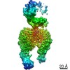
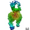
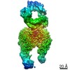
| Title | Cryo EM map of the Gaalphaq-PLCbeta3 complex. | |||||||||
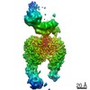
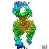
 Map data Map data | Reconstruction of the PLCbeta3 distal CTD in complex with the N-terminal helix of Galphaq | |||||||||
 Sample Sample |
| |||||||||
 Keywords Keywords | GTP-binding protein alpha subunits / phospholipase C beta / coiled-coil domain / PH DOMAIN / EF HAND / C2 DOMAIN / TIM BARREL DOMAIN / phospholipase / GTP hydrolysis / G-protein signaling / membrane targeting / lipase / hydrolase / calcium binding / GTP binding / phospholipids / GTP-BINDING PROTEIN-HYDROLASE complex | |||||||||
| Function / homology | : / Phosphatidylinositol-4, 5-bisphosphate phosphodiesterase beta / G-protein alpha subunit, group Q / G protein-coupled receptor signaling pathway Function and homology information Function and homology information | |||||||||
| Biological species |   Homo sapiens (human) Homo sapiens (human) | |||||||||
| Method | single particle reconstruction / cryo EM / Resolution: 19.0 Å | |||||||||
 Authors Authors | Dutta S / Lyon AM / Tesmer JJG / Skiniotis G | |||||||||
 Citation Citation | Journal: Nat Struct Mol Biol / Year: 2013 Title: Full-length Gα(q)-phospholipase C-β3 structure reveals interfaces of the C-terminal coiled-coil domain. Authors: Angeline M Lyon / Somnath Dutta / Cassandra A Boguth / Georgios Skiniotis / John J G Tesmer /  Abstract: Phospholipase C-β (PLCβ) is directly activated by Gαq, but the molecular basis for how its distal C-terminal domain (CTD) contributes to maximal activity is poorly understood. Herein we present ...Phospholipase C-β (PLCβ) is directly activated by Gαq, but the molecular basis for how its distal C-terminal domain (CTD) contributes to maximal activity is poorly understood. Herein we present both the crystal structure and cryo-EM three-dimensional reconstructions of human full-length PLCβ3 in complex with mouse Gαq. The distal CTD forms an extended monomeric helical bundle consisting of three antiparallel segments with structural similarity to membrane-binding bin-amphiphysin-Rvs (BAR) domains. Sequence conservation of the distal CTD suggests putative membrane and protein interaction sites, the latter of which bind the N-terminal helix of Gαq in both the crystal structure and cryo-EM reconstructions. Functional analysis suggests that the distal CTD has roles in membrane targeting and in optimizing the orientation of the catalytic core at the membrane for maximal rates of lipid hydrolysis. | |||||||||
| History |
|
- Structure visualization
| Movie |
Movie viewer |
|---|---|
| Structure viewer | EM map: SurfViewMolmilJmol/JSmol |
| Supplemental images |

- Downloads & links
-EMDB archive
| Map data | emd_5561.map.gz | 957 KB | EMDB map data format | |
|---|---|---|---|---|
| Header (meta data) | emd-5561-v30.xmlemd-5561.xml | 12.2 KB 12.2 KB | Display Display | EMDB header |
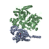
| Images |  emd_5561_1.jpg emd_5561_1.jpg | 34.4 KB | ||
| Archive directory |  http://ftp.pdbj.org/pub/emdb/structures/EMD-5561ftp://ftp.pdbj.org/pub/emdb/structures/EMD-5561 http://ftp.pdbj.org/pub/emdb/structures/EMD-5561ftp://ftp.pdbj.org/pub/emdb/structures/EMD-5561 | HTTPS FTP |
-Related structure data

-Links
| EMDB pages | EMDB (EBI/PDBe) / EMDataResource |
|---|
-Map
| File | Download / File: emd_5561.map.gz / Format: CCP4 / Size: 1001 KB / Type: IMAGE STORED AS FLOATING POINT NUMBER (4 BYTES) | ||||||||||||||||||||||||||||||||||||||||||||||||||||||||||||||||||||
|---|---|---|---|---|---|---|---|---|---|---|---|---|---|---|---|---|---|---|---|---|---|---|---|---|---|---|---|---|---|---|---|---|---|---|---|---|---|---|---|---|---|---|---|---|---|---|---|---|---|---|---|---|---|---|---|---|---|---|---|---|---|---|---|---|---|---|---|---|---|
| Annotation | Reconstruction of the PLCbeta3 distal CTD in complex with the N-terminal helix of Galphaq | ||||||||||||||||||||||||||||||||||||||||||||||||||||||||||||||||||||
| Projections & slices | Image control
Images are generated by Spider. | ||||||||||||||||||||||||||||||||||||||||||||||||||||||||||||||||||||
| Voxel size | X=Y=Z: 4.48 Å | ||||||||||||||||||||||||||||||||||||||||||||||||||||||||||||||||||||
| Density |
| ||||||||||||||||||||||||||||||||||||||||||||||||||||||||||||||||||||
| Symmetry | Space group: 1 | ||||||||||||||||||||||||||||||||||||||||||||||||||||||||||||||||||||
| Details | EMDB XML:
CCP4 map header:
| ||||||||||||||||||||||||||||||||||||||||||||||||||||||||||||||||||||
 Z (Sec.)
 Y (Row.)
 X (Col.)


-Supplemental data
- Sample components
-Entire : Reconstruction of the PLCbeta3 distal CTD in complex with the N-t...
| Entire | Name: Reconstruction of the PLCbeta3 distal CTD in complex with the N-terminal helix of Galphaq |
|---|---|
| Components |
|
-Supramolecule #1000: Reconstruction of the PLCbeta3 distal CTD in complex with the N-t...
| Supramolecule | Name: Reconstruction of the PLCbeta3 distal CTD in complex with the N-terminal helix of Galphaq type: sample / ID: 1000 Details: The sample was monodisperse and had a molecular weight consistent with the theoretical weight. Oligomeric state: Galphaq-PLCb3 dimer / Number unique components: 3 |
|---|---|
| Molecular weight | Experimental: 1.8 MDa / Theoretical: 1.8 MDa Method: Size exclusion chromatography and multi-angle light scattering |
-Macromolecule #1: G alpha q
| Macromolecule | Name: G alpha q / type: protein_or_peptide / ID: 1 / Name.synonym: G a q / Number of copies: 1 / Oligomeric state: monomer / Recombinant expression: Yes |
|---|---|
| Source (natural) | Organism: |
| Molecular weight | Experimental: 44 KDa / Theoretical: 44 KDa |
| Recombinant expression | Organism:  Trichoplusia ni (cabbage looper) / Recombinant plasmid: pFastBacHTA Trichoplusia ni (cabbage looper) / Recombinant plasmid: pFastBacHTA |
| Sequence | GO: GO: 0007202 / InterPro: G-protein alpha subunit, group Q |
-Macromolecule #2: phospholipase C beta 3
| Macromolecule | Name: phospholipase C beta 3 / type: protein_or_peptide / ID: 2 / Name.synonym: PLCbeta3 / Number of copies: 1 / Oligomeric state: monomer / Recombinant expression: Yes |
|---|---|
| Source (natural) | Organism: Homo sapiens (human) / synonym: Human / Location in cell: cytosol and plasma membrane |
| Molecular weight | Experimental: 139 KDa / Theoretical: 139 KDa |
| Recombinant expression | Organism: Trichoplusia ni (cabbage looper) / Recombinant plasmid: pFastBacDual |
| Sequence | GO: G protein-coupled receptor signaling pathway InterPro: Phosphatidylinositol-4, 5-bisphosphate phosphodiesterase beta |
-Experimental details
-Structure determination
| Method | cryo EM |
|---|---|
 Processing Processing | single particle reconstruction |
| Aggregation state | particle |
-Sample preparation
| Concentration | 1 mg/mL |
|---|---|
| Buffer | pH: 8 Details: 20 mM HEPES, pH 8, 200 mM NaCl, 2 mM DTT, 0.9 mM CaCl2, 50 uM GDP, 30 uM AlCl3, 10 mM NaF, 5 mM MgCl2 |
| Grid | Details: glow-discharged Quantifoil R2/200 mesh grid |
| Vitrification | Cryogen name: ETHANE / Chamber humidity: 100 % / Instrument: FEI VITROBOT MARK IV Details: Vitrification carried out in liquid nitrogen atmosphere. Method: Blot 3 microliter sample for 2 seconds. |
- Electron microscopy
| Microscope | FEI TECNAI F20 |
|---|---|
| Temperature | Min: 89 K / Max: 95 K / Average: 89 K |
| Alignment procedure | Legacy - Astigmatism: Objective lens astigmatism was corrected at 135,000 times magnification. |
| Date | Oct 20, 2011 |
| Image recording | Category: CCD / Film or detector model: GATAN ULTRASCAN 4000 (4k x 4k) / Number real images: 350 / Average electron dose: 20 e/Å2 |
| Electron beam | Acceleration voltage: 120 kV / Electron source:  FIELD EMISSION GUN FIELD EMISSION GUN |
| Electron optics | Calibrated magnification: 71138 / Illumination mode: FLOOD BEAM / Imaging mode: BRIGHT FIELD / Cs: 2 mm / Nominal defocus max: 3.5 µm / Nominal defocus min: 1.5 µm / Nominal magnification: 50000 |
| Sample stage | Specimen holder: Used two holders. / Specimen holder model: OTHER |
| Experimental equipment |  Model: Tecnai F20 / Image courtesy: FEI Company |
-Image processing
| CTF correction | Details: each particle |
|---|---|
| Final reconstruction | Algorithm: OTHER / Resolution.type: BY AUTHOR / Resolution: 19.0 Å / Resolution method: FSC 0.5 CUT-OFF / Software - Name: EMAN1 / Number images used: 12692 |
-Atomic model buiding 1
| Initial model | PDB ID: |
|---|---|
| Refinement | Space: REAL |
