 Movie
Movie Controller


+ Open data

- Basic information
| Entry | Database: PDB / ID: 7sg0 | ||||||
|---|---|---|---|---|---|---|---|
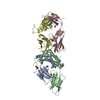
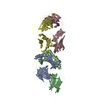
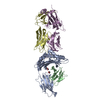
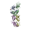
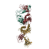
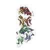
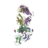
| Title | W316 TCR in complex with HLA-DQ2-omega1 | ||||||
 Components Components |
| ||||||
 Keywords Keywords | IMMUNE SYSTEM / TCR-pHLA complex / Celiac disease | ||||||
| Function / homology |  Function and homology information Function and homology informationMHC class II receptor activity / antigen processing and presentation of peptide or polysaccharide antigen via MHC class II / transport vesicle membrane / Translocation of ZAP-70 to Immunological synapse / Phosphorylation of CD3 and TCR zeta chains / Generation of second messenger molecules / Co-inhibition by PD-1 / MHC class II antigen presentation / trans-Golgi network membrane / peptide antigen assembly with MHC class II protein complex ...MHC class II receptor activity / antigen processing and presentation of peptide or polysaccharide antigen via MHC class II / transport vesicle membrane / Translocation of ZAP-70 to Immunological synapse / Phosphorylation of CD3 and TCR zeta chains / Generation of second messenger molecules / Co-inhibition by PD-1 / MHC class II antigen presentation / trans-Golgi network membrane / peptide antigen assembly with MHC class II protein complex / lumenal side of endoplasmic reticulum membrane / MHC class II protein complex / clathrin-coated endocytic vesicle membrane / ER to Golgi transport vesicle membrane / antigen processing and presentation of exogenous peptide antigen via MHC class II / positive regulation of immune response / peptide antigen binding / positive regulation of T cell activation / Interferon gamma signaling / MHC class II protein complex binding / endocytic vesicle membrane / late endosome membrane / Downstream TCR signaling / adaptive immune response / endosome membrane / immune response / Golgi membrane / lysosomal membrane / membrane / plasma membrane Similarity search - Function | ||||||
| Biological species |  Homo sapiens (human) Homo sapiens (human) | ||||||
| Method |  X-RAY DIFFRACTION / SYNCHROTRON / MOLECULAR REPLACEMENT / Resolution: 3 Å X-RAY DIFFRACTION / SYNCHROTRON / MOLECULAR REPLACEMENT / Resolution: 3 Å | ||||||
 Authors Authors | Ciacchi, L. / Farenc, C. / Petersen, J. / Reid, H.H. / Rossjohn, J. | ||||||
| Funding support |  Australia, 1items Australia, 1items
| ||||||
 Citation Citation | Journal: J.Biol.Chem. / Year: 2022 Title: Structural basis of T cell receptor specificity and cross-reactivity of two HLA-DQ2.5-restricted gluten epitopes in celiac disease. Authors: Ciacchi, L. / Farenc, C. / Dahal-Koirala, S. / Petersen, J. / Sollid, L.M. / Reid, H.H. / Rossjohn, J. | ||||||
| History |
|
- Structure visualization
| Structure viewer | Molecule: MolmilJmol/JSmol |
|---|
- Downloads & links
-Download
| PDBx/mmCIF format | 7sg0.cif.gz | 335.9 KB | Display | PDBx/mmCIF format |
|---|---|---|---|---|
| PDB format | pdb7sg0.ent.gz | 270.4 KB | Display | PDB format |
| PDBx/mmJSON format | 7sg0.json.gz | Tree view | PDBx/mmJSON format | |
| Others |  Other downloads Other downloads |
-Validation report
| Summary document | 7sg0_validation.pdf.gz | 486.9 KB | Display | wwPDB validaton report |
|---|---|---|---|---|
| Full document | 7sg0_full_validation.pdf.gz | 490.6 KB | Display | |
| Data in XML | 7sg0_validation.xml.gz | 28.2 KB | Display | |
| Data in CIF | 7sg0_validation.cif.gz | 37.8 KB | Display | |
| Arichive directory | https://data.pdbj.org/pub/pdb/validation_reports/sg/7sg0ftp://data.pdbj.org/pub/pdb/validation_reports/sg/7sg0 | HTTPS FTP |
-Related structure data
| Related structure data |  7sg1C  7sg2C 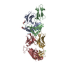 4ozhS 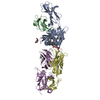 6cugS  6mffS S: Starting model for refinement C: citing same article ( |
|---|---|
| Similar structure data |


-Links
 PDBj
PDBj


- Assembly
| Deposited unit | 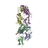
| ||||||||
|---|---|---|---|---|---|---|---|---|---|
| 1 |
| ||||||||
| Unit cell |
|
-Components
-Protein , 2 types, 2 molecules AB
| #1: Protein | Mass: 20683.141 Da / Num. of mol.: 1 Source method: isolated from a genetically manipulated source Source: (gene. exp.) Homo sapiens (human) / Gene: HLA-DQA1 / Cell line (production host): High Five / Production host:  Trichoplusia ni (cabbage looper) / References: UniProt: P01909 Trichoplusia ni (cabbage looper) / References: UniProt: P01909 |
|---|---|
| #2: Protein | Mass: 23700.586 Da / Num. of mol.: 1 Source method: isolated from a genetically manipulated source Source: (gene. exp.) Homo sapiens (human) / Gene: HLA-DQB1 / Cell line (production host): High Five / Production host: Trichoplusia ni (cabbage looper) / References: UniProt: O19712 |
-T-cell receptor, w316, ... , 2 types, 2 molecules DE
| #4: Protein | Mass: 22880.271 Da / Num. of mol.: 1 Source method: isolated from a genetically manipulated source Source: (gene. exp.) Homo sapiens (human)Production host:  |
|---|---|
| #5: Protein | Mass: 26937.852 Da / Num. of mol.: 1 Source method: isolated from a genetically manipulated source Source: (gene. exp.) Homo sapiens (human)Production host: |
-Protein/peptide / Sugars / Non-polymers , 3 types, 6 molecules C

| #3: Protein/peptide | Mass: 1311.437 Da / Num. of mol.: 1 Source method: isolated from a genetically manipulated source Source: (gene. exp.) Homo sapiens (human) / Cell line (production host): High Five / Production host: Trichoplusia ni (cabbage looper) | ||
|---|---|---|---|
| #6: Sugar |  Type: D-saccharide, beta linking / Mass: 221.208 Da / Num. of mol.: 2 / Source method: obtained synthetically / Formula: C8H15NO6 Type: D-saccharide, beta linking / Mass: 221.208 Da / Num. of mol.: 2 / Source method: obtained synthetically / Formula: C8H15NO6#7: Chemical |  Mass: 62.068 Da / Num. of mol.: 3 / Source method: obtained synthetically / Formula: C2H6O2 Mass: 62.068 Da / Num. of mol.: 3 / Source method: obtained synthetically / Formula: C2H6O2 |
-Details
| Has ligand of interest | N |
|---|---|
| Has protein modification | Y |
-Experimental details
-Experiment
| Experiment | Method: X-RAY DIFFRACTION / Number of used crystals: 1 |
|---|
- Sample preparation
| Crystal | Density Matthews: 2.77 Å3/Da / Density % sol: 55.6 % |
|---|---|
| Crystal grow | Temperature: 293.15 K / Method: vapor diffusion, hanging drop Details: 24% PEG3350, 0.25 M (NH4)2SO4 and 0.1 M Tris/HCl at pH 8.0 |
-Data collection
| Diffraction | Mean temperature: 100 K / Serial crystal experiment: N |
|---|---|
| Diffraction source | Source: SYNCHROTRON / Site: Australian Synchrotron / Beamline: MX2 / Wavelength: 0.9537 Å |
| Detector | Type: DECTRIS EIGER X 16M / Detector: PIXEL / Date: Nov 16, 2020 |
| Radiation | Monochromator: M / Protocol: SINGLE WAVELENGTH / Monochromatic (M) / Laue (L): M / Scattering type: x-ray |
| Radiation wavelength | Wavelength: 0.9537 Å / Relative weight: 1 |
| Reflection | Resolution: 3→48.52 Å / Num. obs: 22108 / % possible obs: 99.8 % / Redundancy: 2 % / CC1/2: 0.919 / Net I/σ(I): 5.3 |
| Reflection shell | Resolution: 3→3.1 Å / Redundancy: 2 % / Mean I/σ(I) obs: 1.7 / Num. unique obs: 2177 / CC1/2: 0.384 |
- Processing
| Software |
| ||||||||||||||||||||||||||||||||||||||||||||||||||||||
|---|---|---|---|---|---|---|---|---|---|---|---|---|---|---|---|---|---|---|---|---|---|---|---|---|---|---|---|---|---|---|---|---|---|---|---|---|---|---|---|---|---|---|---|---|---|---|---|---|---|---|---|---|---|---|---|
| Refinement | Method to determine structure: MOLECULAR REPLACEMENT Starting model: 6MFF, 6CUG, 4OZH Resolution: 3→48.52 Å / SU ML: 0.37 / Cross valid method: THROUGHOUT / σ(F): 1.33 / Phase error: 22.75 / Stereochemistry target values: ML
| ||||||||||||||||||||||||||||||||||||||||||||||||||||||
| Solvent computation | Shrinkage radii: 0.9 Å / VDW probe radii: 1.11 Å / Solvent model: FLAT BULK SOLVENT MODEL | ||||||||||||||||||||||||||||||||||||||||||||||||||||||
| Displacement parameters | Biso max: 154.86 Å2 / Biso mean: 55.2196 Å2 / Biso min: 20 Å2 | ||||||||||||||||||||||||||||||||||||||||||||||||||||||
| Refinement step | Cycle: final / Resolution: 3→48.52 Å
| ||||||||||||||||||||||||||||||||||||||||||||||||||||||
| LS refinement shell | Refine-ID: X-RAY DIFFRACTION / Rfactor Rfree error: 0 / Total num. of bins used: 8 / % reflection obs: 100 %
| ||||||||||||||||||||||||||||||||||||||||||||||||||||||
| Refinement TLS params. | Method: refined / Origin x: 63.2724 Å / Origin y: -71.0908 Å / Origin z: 21.0647 Å
| ||||||||||||||||||||||||||||||||||||||||||||||||||||||
| Refinement TLS group |
|
