 Movie
Movie Controller

[English] 日本語
 Yorodumi
- PDB-2ian: Structural basis for recognition of mutant self by a tumor-specif... -
+ Open data

- Basic information
| Entry | Database: PDB / ID: 2ian | ||||||
|---|---|---|---|---|---|---|---|
| Title | Structural basis for recognition of mutant self by a tumor-specific, MHC class II-restricted TCR | ||||||
 Components Components |
| ||||||
 Keywords Keywords | IMMUNE SYSTEM / major histocompatibility complex / T cell receptor / T cell stimulation / melanoma / tumor antigen | ||||||
| Function / homology |  Function and homology information Function and homology informationmethylglyoxal biosynthetic process / methylglyoxal synthase / methylglyoxal synthase activity / regulation of interleukin-4 production / regulation of interleukin-10 production / autolysosome membrane / myeloid dendritic cell antigen processing and presentation / antigen processing and presentation of endogenous peptide antigen via MHC class II / regulation of T-helper cell differentiation / positive regulation of CD4-positive, CD25-positive, alpha-beta regulatory T cell differentiation ...methylglyoxal biosynthetic process / methylglyoxal synthase / methylglyoxal synthase activity / regulation of interleukin-4 production / regulation of interleukin-10 production / autolysosome membrane / myeloid dendritic cell antigen processing and presentation / antigen processing and presentation of endogenous peptide antigen via MHC class II / regulation of T-helper cell differentiation / positive regulation of CD4-positive, CD25-positive, alpha-beta regulatory T cell differentiation / positive regulation of CD4-positive, alpha-beta T cell activation / antigen processing and presentation of peptide or polysaccharide antigen via MHC class II / positive regulation of T cell mediated immune response to tumor cell / triose-phosphate isomerase / positive regulation of memory T cell differentiation / triose-phosphate isomerase activity / positive regulation of monocyte differentiation / inflammatory response to antigenic stimulus / CD4 receptor binding / Gluconeogenesis / glyceraldehyde-3-phosphate biosynthetic process / glycerol catabolic process / Glycolysis / alpha-beta T cell receptor complex / T-helper 1 type immune response / intermediate filament / canonical glycolysis / transport vesicle membrane / Translocation of ZAP-70 to Immunological synapse / Phosphorylation of CD3 and TCR zeta chains / polysaccharide binding / negative regulation of type II interferon production / humoral immune response / alpha-beta T cell activation / Generation of second messenger molecules / macrophage differentiation / immunological synapse / Co-inhibition by PD-1 / epidermis development / detection of bacterium / negative regulation of T cell proliferation / T cell receptor binding / MHC class II antigen presentation / positive regulation of insulin secretion involved in cellular response to glucose stimulus / trans-Golgi network membrane / glycolytic process / gluconeogenesis / response to bacterium / lumenal side of endoplasmic reticulum membrane / protein tetramerization / ER to Golgi transport vesicle membrane / negative regulation of inflammatory response to antigenic stimulus / clathrin-coated endocytic vesicle membrane / peptide antigen assembly with MHC class II protein complex / MHC class II protein complex / positive regulation of T cell mediated cytotoxicity / structural constituent of cytoskeleton / antigen processing and presentation of exogenous peptide antigen via MHC class II / positive regulation of immune response / peptide antigen binding / cognition / positive regulation of T cell activation / Interferon gamma signaling / Immunoregulatory interactions between a Lymphoid and a non-Lymphoid cell / endocytic vesicle membrane / MHC class II protein complex binding / late endosome membrane / Downstream TCR signaling / T cell receptor signaling pathway / early endosome membrane / adaptive immune response / lysosome / immune response / Golgi membrane / external side of plasma membrane / lysosomal membrane / ubiquitin protein ligase binding / cell surface / signal transduction / protein homodimerization activity / : / extracellular exosome / membrane / nucleus / plasma membrane / cytosol Similarity search - Function | ||||||
| Biological species |  Homo sapiens (human) Homo sapiens (human) | ||||||
| Method |  X-RAY DIFFRACTION / MOLECULAR REPLACEMENT / Resolution: 2.8 Å X-RAY DIFFRACTION / MOLECULAR REPLACEMENT / Resolution: 2.8 Å | ||||||
 Authors Authors | Deng, L. / Langley, R.J. / Mariuzza, R.A. | ||||||
 Citation Citation | Journal: Nat.Immunol. / Year: 2007 Title: Structural basis for the recognition of mutant self by a tumor-specific, MHC class II-restricted T cell receptor Authors: Deng, L. / Langley, R.J. / Brown, P.H. / Xu, G. / Teng, L. / Wang, Q. / Gonzales, M.I. / Callender, G.G. / Nishimura, M.I. / Topalian, S.L. / Mariuzza, R.A. | ||||||
| History |
|
- Structure visualization
| Structure viewer | Molecule: MolmilJmol/JSmol |
|---|
- Downloads & links
-Download
| PDBx/mmCIF format | 2ian.cif.gz | 627.7 KB | Display | PDBx/mmCIF format |
|---|---|---|---|---|
| PDB format | pdb2ian.ent.gz | 516.5 KB | Display | PDB format |
| PDBx/mmJSON format | 2ian.json.gz | Tree view | PDBx/mmJSON format | |
| Others |  Other downloads Other downloads |
-Validation report
| Arichive directory | https://data.pdbj.org/pub/pdb/validation_reports/ia/2ianftp://data.pdbj.org/pub/pdb/validation_reports/ia/2ian | HTTPS FTP |
|---|
-Related structure data
| Related structure data |  2ialC  2iamC 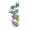 1fytS 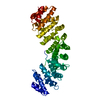 1ialS C: citing same article ( S: Starting model for refinement |
|---|---|
| Similar structure data |


-Links
 PDBj
PDBj


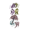
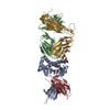
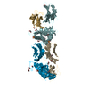
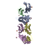
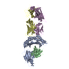
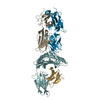
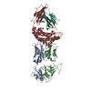
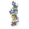
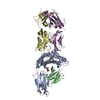
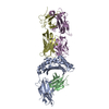
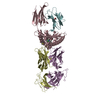
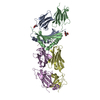
- Assembly
| Deposited unit | 
| ||||||||
|---|---|---|---|---|---|---|---|---|---|
| 1 | 
| ||||||||
| 2 | 
| ||||||||
| 3 | 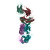
| ||||||||
| 4 | 
| ||||||||
| Unit cell |
| ||||||||
| Details | Each aymmetric unit contains four biological units. |
-Components
-HLA class II histocompatibility antigen, ... , 2 types, 8 molecules AFKPBGLQ
| #1: Protein | Mass: 21155.904 Da / Num. of mol.: 4 / Fragment: residues 1-182 (26-207) Source method: isolated from a genetically manipulated source Source: (gene. exp.) Homo sapiens (human) / Gene: HLA-DRA / Plasmid: pT7-7 / Species (production host): Escherichia coli / Production host:  #2: Protein | Mass: 22080.664 Da / Num. of mol.: 4 / Fragment: residues 1-190 (30-219) Source method: isolated from a genetically manipulated source Source: (gene. exp.) Homo sapiens (human) / Gene: HLA-DRB1 / Plasmid: pT7-7 / Species (production host): Escherichia coli / Production host: |
|---|
-CD4+ T cell receptor E8 ... , 2 types, 8 molecules DINSEJOT
| #4: Protein | Mass: 22399.682 Da / Num. of mol.: 4 / Mutation: T156C Source method: isolated from a genetically manipulated source Source: (gene. exp.) Homo sapiens (human) / Plasmid: pT7-7 / Species (production host): Escherichia coli / Production host: #5: Protein | Mass: 27088.197 Da / Num. of mol.: 4 / Mutation: S167C Source method: isolated from a genetically manipulated source Source: (gene. exp.) Homo sapiens (human) / Plasmid: pT7-7 / Species (production host): Escherichia coli / Production host: |
|---|
-Protein/peptide / Non-polymers , 2 types, 80 molecules CHMR
| #3: Protein/peptide | Mass: 1469.659 Da / Num. of mol.: 4 / Fragment: residues 23-37 (22-36) Source method: isolated from a genetically manipulated source Source: (gene. exp.) Homo sapiens (human) / Gene: TPI1, TPI / Plasmid: pT7-7 / Species (production host): Escherichia coli / Production host: #6: Water | ChemComp-HOH / | Mass: 18.015 Da / Num. of mol.: 76 / Source method: isolated from a natural source / Formula: H2O |
|---|
-Details
| Has protein modification | Y |
|---|
-Experimental details
-Experiment
| Experiment | Method: X-RAY DIFFRACTION / Number of used crystals: 1 |
|---|
- Sample preparation
| Crystal | Density Matthews: 2.95 Å3/Da / Density % sol: 58.33 % |
|---|---|
| Crystal grow | Temperature: 295 K / Method: vapor diffusion, hanging drop / pH: 8.5 Details: 10% PEG6000, 0.1M di-ammonium phosphate, 0.1M Tris, pH 8.5, VAPOR DIFFUSION, HANGING DROP, temperature 295K |
-Data collection
| Diffraction | Mean temperature: 100 K |
|---|---|
| Diffraction source | Source: ROTATING ANODE / Type: RIGAKU / Wavelength: 1.54 Å |
| Detector | Type: RIGAKU RAXIS IV / Detector: IMAGE PLATE / Date: Aug 25, 2005 |
| Radiation | Protocol: SINGLE WAVELENGTH / Monochromatic (M) / Laue (L): M / Scattering type: x-ray |
| Radiation wavelength | Wavelength: 1.54 Å / Relative weight: 1 |
| Reflection | Resolution: 2.8→50 Å / Num. obs: 100306 / % possible obs: 93.8 % / Rmerge(I) obs: 0.082 / Net I/σ(I): 11.1 |
| Reflection shell | Resolution: 2.8→2.94 Å / Rmerge(I) obs: 0.235 / Mean I/σ(I) obs: 3.9 / Num. unique all: 18951 / % possible all: 92.8 |
- Processing
| Software |
| ||||||||||||||||||||||||||||||||||||||||||||||||||||||||||||||||||||||||||||||||||||||||||||||||||||||||||||||||||||||||||||||||||||||||||||||||||||||||||||||||||||||||||
|---|---|---|---|---|---|---|---|---|---|---|---|---|---|---|---|---|---|---|---|---|---|---|---|---|---|---|---|---|---|---|---|---|---|---|---|---|---|---|---|---|---|---|---|---|---|---|---|---|---|---|---|---|---|---|---|---|---|---|---|---|---|---|---|---|---|---|---|---|---|---|---|---|---|---|---|---|---|---|---|---|---|---|---|---|---|---|---|---|---|---|---|---|---|---|---|---|---|---|---|---|---|---|---|---|---|---|---|---|---|---|---|---|---|---|---|---|---|---|---|---|---|---|---|---|---|---|---|---|---|---|---|---|---|---|---|---|---|---|---|---|---|---|---|---|---|---|---|---|---|---|---|---|---|---|---|---|---|---|---|---|---|---|---|---|---|---|---|---|---|---|---|
| Refinement | Method to determine structure: MOLECULAR REPLACEMENT Starting model: 1FYT, 1IAL Resolution: 2.8→45.45 Å / Cor.coef. Fo:Fc: 0.906 / Cor.coef. Fo:Fc free: 0.843 / SU B: 28.895 / SU ML: 0.308 / Cross valid method: THROUGHOUT / ESU R: 0.91 / ESU R Free: 0.386 / Stereochemistry target values: MAXIMUM LIKELIHOOD
| ||||||||||||||||||||||||||||||||||||||||||||||||||||||||||||||||||||||||||||||||||||||||||||||||||||||||||||||||||||||||||||||||||||||||||||||||||||||||||||||||||||||||||
| Solvent computation | Ion probe radii: 0.8 Å / Shrinkage radii: 0.8 Å / VDW probe radii: 1.2 Å / Solvent model: MASK | ||||||||||||||||||||||||||||||||||||||||||||||||||||||||||||||||||||||||||||||||||||||||||||||||||||||||||||||||||||||||||||||||||||||||||||||||||||||||||||||||||||||||||
| Displacement parameters | Biso mean: 14.606 Å2
| ||||||||||||||||||||||||||||||||||||||||||||||||||||||||||||||||||||||||||||||||||||||||||||||||||||||||||||||||||||||||||||||||||||||||||||||||||||||||||||||||||||||||||
| Refinement step | Cycle: LAST / Resolution: 2.8→45.45 Å
| ||||||||||||||||||||||||||||||||||||||||||||||||||||||||||||||||||||||||||||||||||||||||||||||||||||||||||||||||||||||||||||||||||||||||||||||||||||||||||||||||||||||||||
| Refine LS restraints |
| ||||||||||||||||||||||||||||||||||||||||||||||||||||||||||||||||||||||||||||||||||||||||||||||||||||||||||||||||||||||||||||||||||||||||||||||||||||||||||||||||||||||||||
| LS refinement shell | Resolution: 2.8→2.873 Å / Total num. of bins used: 20
|
