 Movie
Movie Controller

[English] 日本語
 Yorodumi
- PDB-2ial: Structural basis for recognition of mutant self by a tumor-specif... -
+ Open data

- Basic information
| Entry | Database: PDB / ID: 2ial | ||||||
|---|---|---|---|---|---|---|---|
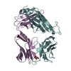
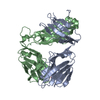
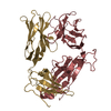
| Title | Structural basis for recognition of mutant self by a tumor-specific, MHC class II-restricted TCR | ||||||
 Components Components |
| ||||||
 Keywords Keywords | IMMUNE SYSTEM / major histocompatibility complex / T cell receptor / T cell stimulation / melanoma / tumor antigen | ||||||
| Function / homology |  Function and homology information Function and homology informationalpha-beta T cell receptor complex / Translocation of ZAP-70 to Immunological synapse / Phosphorylation of CD3 and TCR zeta chains / alpha-beta T cell activation / Generation of second messenger molecules / Co-inhibition by PD-1 / response to bacterium / Immunoregulatory interactions between a Lymphoid and a non-Lymphoid cell / T cell receptor signaling pathway / Downstream TCR signaling ...alpha-beta T cell receptor complex / Translocation of ZAP-70 to Immunological synapse / Phosphorylation of CD3 and TCR zeta chains / alpha-beta T cell activation / Generation of second messenger molecules / Co-inhibition by PD-1 / response to bacterium / Immunoregulatory interactions between a Lymphoid and a non-Lymphoid cell / T cell receptor signaling pathway / Downstream TCR signaling / adaptive immune response / immune response / membrane / plasma membrane Similarity search - Function | ||||||
| Biological species |  Homo sapiens (human) Homo sapiens (human) | ||||||
| Method |  X-RAY DIFFRACTION / SYNCHROTRON / MOLECULAR REPLACEMENT / Resolution: 1.92 Å X-RAY DIFFRACTION / SYNCHROTRON / MOLECULAR REPLACEMENT / Resolution: 1.92 Å | ||||||
 Authors Authors | Deng, L. / Langley, R.J. / Mariuzza, R.A. | ||||||
 Citation Citation | Journal: Nat.Immunol. / Year: 2007 Title: Structural basis for the recognition of mutant self by a tumor-specific, MHC class II-restricted T cell receptor Authors: Deng, L. / Langley, R.J. / Brown, P.H. / Xu, G. / Teng, L. / Wang, Q. / Gonzales, M.I. / Callender, G.G. / Nishimura, M.I. / Topalian, S.L. / Mariuzza, R.A. | ||||||
| History |
|
- Structure visualization
| Structure viewer | Molecule: MolmilJmol/JSmol |
|---|
- Downloads & links
-Download
| PDBx/mmCIF format | 2ial.cif.gz | 184.3 KB | Display | PDBx/mmCIF format |
|---|---|---|---|---|
| PDB format | pdb2ial.ent.gz | 148.1 KB | Display | PDB format |
| PDBx/mmJSON format | 2ial.json.gz | Tree view | PDBx/mmJSON format | |
| Others |  Other downloads Other downloads |
-Validation report
| Arichive directory | https://data.pdbj.org/pub/pdb/validation_reports/ia/2ialftp://data.pdbj.org/pub/pdb/validation_reports/ia/2ial | HTTPS FTP |
|---|
-Related structure data
| Related structure data |  2iamC  2ianC 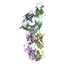 1fytS C: citing same article ( S: Starting model for refinement |
|---|---|
| Similar structure data |


-Links
 PDBj
PDBj


- Assembly
| Deposited unit | 
| ||||||||
|---|---|---|---|---|---|---|---|---|---|
| 1 | 
| ||||||||
| 2 | 
| ||||||||
| Unit cell |
| ||||||||
| Details | Each asymmetric unit contains two biological units. |
-Components
| #1: Protein | Mass: 22399.682 Da / Num. of mol.: 2 / Mutation: T156C Source method: isolated from a genetically manipulated source Source: (gene. exp.) Homo sapiens (human) / Plasmid: pT7-7 / Species (production host): Escherichia coli / Production host:  #2: Protein | Mass: 27088.197 Da / Num. of mol.: 2 / Mutation: S167C Source method: isolated from a genetically manipulated source Source: (gene. exp.) Homo sapiens (human) / Plasmid: pT7-7 / Species (production host): Escherichia coli / Production host: #3: Water | ChemComp-HOH / |  Mass: 18.015 Da / Num. of mol.: 243 / Source method: isolated from a natural source / Formula: H2O Mass: 18.015 Da / Num. of mol.: 243 / Source method: isolated from a natural source / Formula: H2OHas protein modification | Y | |
|---|
-Experimental details
-Experiment
| Experiment | Method: X-RAY DIFFRACTION / Number of used crystals: 1 |
|---|
- Sample preparation
| Crystal | Density Matthews: 2.39 Å3/Da / Density % sol: 48.55 % |
|---|---|
| Crystal grow | Temperature: 295 K / Method: vapor diffusion, hanging drop / pH: 7 Details: 10% PEG-2000 MME, 0.1M Tris, pH 7.0, VAPOR DIFFUSION, HANGING DROP, temperature 295K |
-Data collection
| Diffraction | Mean temperature: 100 K |
|---|---|
| Diffraction source | Source: SYNCHROTRON / Site: NSLS  / Beamline: X29A / Wavelength: 1 Å / Beamline: X29A / Wavelength: 1 Å |
| Detector | Type: ADSC QUANTUM 315 / Detector: CCD / Date: Jul 4, 2005 |
| Radiation | Protocol: SINGLE WAVELENGTH / Monochromatic (M) / Laue (L): M / Scattering type: x-ray |
| Radiation wavelength | Wavelength: 1 Å / Relative weight: 1 |
| Reflection | Resolution: 1.92→50 Å / Num. obs: 71032 / Rmerge(I) obs: 0.108 / Net I/σ(I): 13.1 |
| Reflection shell | Resolution: 1.92→1.99 Å / Rmerge(I) obs: 0.491 / Mean I/σ(I) obs: 1.5 / Num. unique all: 9536 / % possible all: 78.1 |
- Processing
| Software |
| ||||||||||||||||||||||||||||||||||||||||||||||||||||||||||||||||||||||||||||||||||||||||||||||||||||||||||||||||||||||||||||||||||||||||||||||||||||||||||||||||||||||||||
|---|---|---|---|---|---|---|---|---|---|---|---|---|---|---|---|---|---|---|---|---|---|---|---|---|---|---|---|---|---|---|---|---|---|---|---|---|---|---|---|---|---|---|---|---|---|---|---|---|---|---|---|---|---|---|---|---|---|---|---|---|---|---|---|---|---|---|---|---|---|---|---|---|---|---|---|---|---|---|---|---|---|---|---|---|---|---|---|---|---|---|---|---|---|---|---|---|---|---|---|---|---|---|---|---|---|---|---|---|---|---|---|---|---|---|---|---|---|---|---|---|---|---|---|---|---|---|---|---|---|---|---|---|---|---|---|---|---|---|---|---|---|---|---|---|---|---|---|---|---|---|---|---|---|---|---|---|---|---|---|---|---|---|---|---|---|---|---|---|---|---|---|
| Refinement | Method to determine structure: MOLECULAR REPLACEMENT Starting model: 1FYT Resolution: 1.92→44.31 Å / Cor.coef. Fo:Fc: 0.947 / Cor.coef. Fo:Fc free: 0.91 / SU B: 9.901 / SU ML: 0.142 / Cross valid method: THROUGHOUT / ESU R: 0.18 / ESU R Free: 0.173 / Stereochemistry target values: MAXIMUM LIKELIHOOD
| ||||||||||||||||||||||||||||||||||||||||||||||||||||||||||||||||||||||||||||||||||||||||||||||||||||||||||||||||||||||||||||||||||||||||||||||||||||||||||||||||||||||||||
| Solvent computation | Ion probe radii: 0.8 Å / Shrinkage radii: 0.8 Å / VDW probe radii: 1.2 Å / Solvent model: MASK | ||||||||||||||||||||||||||||||||||||||||||||||||||||||||||||||||||||||||||||||||||||||||||||||||||||||||||||||||||||||||||||||||||||||||||||||||||||||||||||||||||||||||||
| Displacement parameters | Biso mean: 31.079 Å2
| ||||||||||||||||||||||||||||||||||||||||||||||||||||||||||||||||||||||||||||||||||||||||||||||||||||||||||||||||||||||||||||||||||||||||||||||||||||||||||||||||||||||||||
| Refinement step | Cycle: LAST / Resolution: 1.92→44.31 Å
| ||||||||||||||||||||||||||||||||||||||||||||||||||||||||||||||||||||||||||||||||||||||||||||||||||||||||||||||||||||||||||||||||||||||||||||||||||||||||||||||||||||||||||
| Refine LS restraints |
| ||||||||||||||||||||||||||||||||||||||||||||||||||||||||||||||||||||||||||||||||||||||||||||||||||||||||||||||||||||||||||||||||||||||||||||||||||||||||||||||||||||||||||
| LS refinement shell | Resolution: 1.918→1.968 Å / Total num. of bins used: 20
|
