 Movie
Movie Controller

[English] 日本語
 Yorodumi
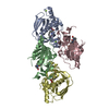
- PDB-1klu: Crystal structure of HLA-DR1/TPI(23-37) complexed with staphyloco... -
+ Open data

- Basic information
| Entry | Database: PDB / ID: 1klu | ||||||
|---|---|---|---|---|---|---|---|
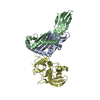
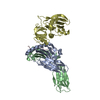
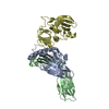
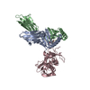
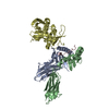
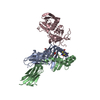
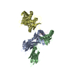
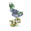
| Title | Crystal structure of HLA-DR1/TPI(23-37) complexed with staphylococcal enterotoxin C3 variant 3B2 (SEC3-3B2) | ||||||
 Components Components |
| ||||||
 Keywords Keywords | IMMUNE SYSTEM/TOXIN / HLA-DR1/TPI / enterotoxin C3 / human melanoma antigen / CD4+ T cells / IMMUNE SYSTEM-TOXIN COMPLEX | ||||||
| Function / homology |  Function and homology information Function and homology informationmethylglyoxal biosynthetic process / methylglyoxal synthase / methylglyoxal synthase activity / regulation of interleukin-4 production / regulation of interleukin-10 production / myeloid dendritic cell antigen processing and presentation / antigen processing and presentation of endogenous peptide antigen via MHC class II / autolysosome membrane / positive regulation of CD4-positive, CD25-positive, alpha-beta regulatory T cell differentiation / regulation of T-helper cell differentiation ...methylglyoxal biosynthetic process / methylglyoxal synthase / methylglyoxal synthase activity / regulation of interleukin-4 production / regulation of interleukin-10 production / myeloid dendritic cell antigen processing and presentation / antigen processing and presentation of endogenous peptide antigen via MHC class II / autolysosome membrane / positive regulation of CD4-positive, CD25-positive, alpha-beta regulatory T cell differentiation / regulation of T-helper cell differentiation / positive regulation of CD4-positive, alpha-beta T cell activation / antigen processing and presentation of peptide or polysaccharide antigen via MHC class II / inflammatory response to antigenic stimulus / positive regulation of T cell mediated immune response to tumor cell / positive regulation of memory T cell differentiation / triose-phosphate isomerase / triose-phosphate isomerase activity / positive regulation of monocyte differentiation / CD4 receptor binding / Gluconeogenesis / glyceraldehyde-3-phosphate biosynthetic process / glycerol catabolic process / Glycolysis / canonical glycolysis / T-helper 1 type immune response / intermediate filament / transport vesicle membrane / Translocation of ZAP-70 to Immunological synapse / humoral immune response / Phosphorylation of CD3 and TCR zeta chains / epidermis development / polysaccharide binding / negative regulation of type II interferon production / macrophage differentiation / Generation of second messenger molecules / immunological synapse / Co-inhibition by PD-1 / detection of bacterium / negative regulation of T cell proliferation / T cell receptor binding / MHC class II antigen presentation / gluconeogenesis / glycolytic process / trans-Golgi network membrane / positive regulation of insulin secretion involved in cellular response to glucose stimulus / lumenal side of endoplasmic reticulum membrane / protein tetramerization / negative regulation of inflammatory response to antigenic stimulus / ER to Golgi transport vesicle membrane / clathrin-coated endocytic vesicle membrane / peptide antigen assembly with MHC class II protein complex / positive regulation of T cell mediated cytotoxicity / MHC class II protein complex / cognition / antigen processing and presentation of exogenous peptide antigen via MHC class II / positive regulation of immune response / peptide antigen binding / positive regulation of T cell activation / Interferon gamma signaling / structural constituent of cytoskeleton / endocytic vesicle membrane / MHC class II protein complex binding / T cell receptor signaling pathway / Downstream TCR signaling / late endosome membrane / toxin activity / early endosome membrane / adaptive immune response / lysosome / immune response / external side of plasma membrane / Golgi membrane / lysosomal membrane / ubiquitin protein ligase binding / cell surface / signal transduction / protein homodimerization activity / : / extracellular exosome / extracellular region / membrane / metal ion binding / nucleus / plasma membrane / cytosol Similarity search - Function | ||||||
| Biological species |  Homo sapiens (human) Homo sapiens (human)  Staphylococcus aureus (bacteria) Staphylococcus aureus (bacteria) | ||||||
| Method |  X-RAY DIFFRACTION / SYNCHROTRON / Resolution: 1.93 Å X-RAY DIFFRACTION / SYNCHROTRON / Resolution: 1.93 Å | ||||||
 Authors Authors | Sundberg, E.J. / Sawicki, M.W. / Andersen, P.S. / Sidney, J. / Sette, A. / Mariuzza, R.A. | ||||||
 Citation Citation | Journal: J.Mol.Biol. / Year: 2002 Title: Minor structural changes in a mutated human melanoma antigen correspond to dramatically enhanced stimulation of a CD4+ tumor-infiltrating lymphocyte line. Authors: Sundberg, E.J. / Sawicki, M.W. / Southwood, S. / Andersen, P.S. / Sette, A. / Mariuzza, R.A. #1: Journal: J.Exp.Med. / Year: 1999Title: Biochemical identification of a mutated human melanoma antigen recognized by CD4+ T cells Authors: Pieper, R. / Christian, R.E. / Gonzales, M.I. / Nishimura, M.I. / Gupta, G. / Settlage, R.E. / Shabanowitz, J. / Rosenberg, S.A. / Hunt, D.F. / Topalian, S.L. | ||||||
| History |
|
- Structure visualization
| Structure viewer | Molecule: MolmilJmol/JSmol |
|---|
- Downloads & links
-Download
| PDBx/mmCIF format | 1klu.cif.gz | 146.8 KB | Display | PDBx/mmCIF format |
|---|---|---|---|---|
| PDB format | pdb1klu.ent.gz | 114.2 KB | Display | PDB format |
| PDBx/mmJSON format | 1klu.json.gz | Tree view | PDBx/mmJSON format | |
| Others |  Other downloads Other downloads |
-Validation report
| Arichive directory | https://data.pdbj.org/pub/pdb/validation_reports/kl/1kluftp://data.pdbj.org/pub/pdb/validation_reports/kl/1klu | HTTPS FTP |
|---|
-Related structure data


-Links
 PDBj
PDBj


- Assembly
| Deposited unit | 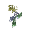
| ||||||||||
|---|---|---|---|---|---|---|---|---|---|---|---|
| 1 |
| ||||||||||
| Unit cell |
|
-Components
| #1: Protein | Mass: 20784.455 Da / Num. of mol.: 1 / Fragment: residues 29-207 Source method: isolated from a genetically manipulated source Source: (gene. exp.) Homo sapiens (human) / Production host: |
|---|---|
| #2: Protein | Mass: 22080.664 Da / Num. of mol.: 1 / Fragment: residues 30-219 Source method: isolated from a genetically manipulated source Source: (gene. exp.) Homo sapiens (human) / Production host: |
| #3: Protein/peptide | Mass: 1469.659 Da / Num. of mol.: 1 / Fragment: residues 23-37 / Source method: obtained synthetically / Details: peptide synthesis / References: UniProt: P60174*PLUS |
| #4: Protein | Mass: 27721.068 Da / Num. of mol.: 1 / Fragment: residues 29-266 Source method: isolated from a genetically manipulated source Source: (gene. exp.) Staphylococcus aureus (bacteria) / Variant: 3B2 / Production host: |
| #5: Water | ChemComp-HOH /  Mass: 18.015 Da / Num. of mol.: 405 / Source method: isolated from a natural source / Formula: H2O Mass: 18.015 Da / Num. of mol.: 405 / Source method: isolated from a natural source / Formula: H2O |
| Has protein modification | Y |
-Experimental details
-Experiment
| Experiment | Method: X-RAY DIFFRACTION / Number of used crystals: 1 |
|---|
- Sample preparation
| Crystal | Density Matthews: 4.74 Å3/Da / Density % sol: 74.05 % | ||||||||||||||||||||||||||||||
|---|---|---|---|---|---|---|---|---|---|---|---|---|---|---|---|---|---|---|---|---|---|---|---|---|---|---|---|---|---|---|---|
| Crystal grow | Temperature: 298 K / Method: vapor diffusion, hanging drop / pH: 5.2 Details: PEG 4000, sodium acetate, ethylene glycol, pH 5.2, VAPOR DIFFUSION, HANGING DROP at 298K, VAPOR DIFFUSION, HANGING DROP | ||||||||||||||||||||||||||||||
| Crystal grow | *PLUS | ||||||||||||||||||||||||||||||
| Components of the solutions | *PLUS
|
-Data collection
| Diffraction | Mean temperature: 100 K |
|---|---|
| Diffraction source | Source: SYNCHROTRON / Site: NSLS  / Beamline: X12C / Wavelength: 0.9785 Å / Beamline: X12C / Wavelength: 0.9785 Å |
| Detector | Type: BRANDEIS - B4 / Detector: CCD / Date: Aug 30, 2001 |
| Radiation | Protocol: SINGLE WAVELENGTH / Monochromatic (M) / Laue (L): M / Scattering type: x-ray |
| Radiation wavelength | Wavelength: 0.9785 Å / Relative weight: 1 |
| Reflection | Resolution: 1.93→100 Å / Num. all: 99361 / % possible obs: 98.1 % / Observed criterion σ(F): 2 / Observed criterion σ(I): 1 / Biso Wilson estimate: 7.8 Å2 / Rsym value: 0.095 |
| Reflection | *PLUS Lowest resolution: 100 Å / Num. obs: 99361 / % possible obs: 99.8 % / Num. measured all: 311680 / Rmerge(I) obs: 0.095 |
| Reflection shell | *PLUS Highest resolution: 1.93 Å / Lowest resolution: 1.96 Å / % possible obs: 98.1 % / Rmerge(I) obs: 0.479 / Mean I/σ(I) obs: 2 |
- Processing
| Software |
| ||||||||||||||||||||
|---|---|---|---|---|---|---|---|---|---|---|---|---|---|---|---|---|---|---|---|---|---|
| Refinement | Resolution: 1.93→14.95 Å / Rfactor Rfree error: 0.003 / Data cutoff high absF: 152684.3 / Data cutoff low absF: 0 / Isotropic thermal model: RESTRAINED / Cross valid method: THROUGHOUT / σ(F): 2
| ||||||||||||||||||||
| Solvent computation | Solvent model: FLAT MODEL / Bsol: 43.6737 Å2 / ksol: 0.37004 e/Å3 | ||||||||||||||||||||
| Displacement parameters | Biso mean: 27.1 Å2
| ||||||||||||||||||||
| Refine analyze |
| ||||||||||||||||||||
| Refinement step | Cycle: LAST / Resolution: 1.93→14.95 Å
| ||||||||||||||||||||
| Refine LS restraints |
| ||||||||||||||||||||
| LS refinement shell | Resolution: 1.93→2.05 Å / Rfactor Rfree error: 0.01 / Total num. of bins used: 6
| ||||||||||||||||||||
| Xplor file |
| ||||||||||||||||||||
| Refinement | *PLUS Num. reflection Rfree: 4240 / % reflection Rfree: 4.3 % / Rfactor Rfree: 0.22 | ||||||||||||||||||||
| Solvent computation | *PLUS | ||||||||||||||||||||
| Displacement parameters | *PLUS | ||||||||||||||||||||
| Refine LS restraints | *PLUS
| ||||||||||||||||||||
| LS refinement shell | *PLUS Lowest resolution: 1.96 Å / Rfactor Rfree: 0.234 / Rfactor Rwork: 0.254 |
