 Movie
Movie Controller

[English] 日本語
 Yorodumi
- PDB-7see: Structure of E. coli LetB delta (Ring6) mutant, Ring1 in the clos... -
+ Open data

- Basic information
| Entry | Database: PDB / ID: 7see | |||||||||||||||
|---|---|---|---|---|---|---|---|---|---|---|---|---|---|---|---|---|
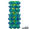
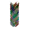
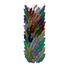
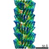
| Title | Structure of E. coli LetB delta (Ring6) mutant, Ring1 in the closed state (Model 1) | |||||||||||||||
 Components Components | MCE family protein, Intermembrane transport protein YebT chimera | |||||||||||||||
 Keywords Keywords | LIPID TRANSPORT / bacterial cell envelope / MCE | |||||||||||||||
| Function / homology |  Function and homology information Function and homology informationintermembrane lipid transfer / membrane organization / outer membrane-bounded periplasmic space / identical protein binding / plasma membrane Similarity search - Function | |||||||||||||||
| Biological species |  | |||||||||||||||
| Method | ELECTRON MICROSCOPY / single particle reconstruction / cryo EM / Resolution: 3.2 Å | |||||||||||||||
 Authors Authors | Vieni, C. / Coudray, N. / Bhabha, G. / Ekiert, D. | |||||||||||||||
| Funding support |  United States, 4items United States, 4items
| |||||||||||||||
 Citation Citation | Journal: J Mol Biol / Year: 2022 Title: Role of Ring6 in the Function of the E. coli MCE Protein LetB. Authors: Casey Vieni / Nicolas Coudray / Georgia L Isom / Gira Bhabha / Damian C Ekiert / Abstract: LetB is a tunnel-forming protein found in the cell envelope of some double-membraned bacteria, and is thought to be important for the transport of lipids between the inner and outer membranes. In ...LetB is a tunnel-forming protein found in the cell envelope of some double-membraned bacteria, and is thought to be important for the transport of lipids between the inner and outer membranes. In Escherichia coli the LetB tunnel is formed from a stack of seven rings (Ring1 - Ring7), in which each ring is composed of a homo-hexameric assembly of MCE domains. The primary sequence of each MCE domain of the LetB protein is substantially divergent from the others, making each MCE ring unique in nature. The role of each MCE domain and how it contributes to the function of LetB is not well understood. Here we probed the importance of each MCE ring for the function of LetB, using a combination of bacterial growth assays and cryo-EM. Surprisingly, we find that ΔRing3 and ΔRing6 mutants, in which Ring3 and Ring6 have been deleted, confer increased resistance to membrane perturbing agents. Specific mutations in the pore-lining loops of Ring6 similarly confer increased resistance. A cryo-EM structure of the ΔRing6 mutant shows that despite the absence of Ring6, which leads to a shorter assembly, the overall architecture is maintained, highlighting the modular nature of MCE proteins. Previous work has shown that Ring6 is dynamic and in its closed state, may restrict the passage of substrate through the tunnel. Our work suggests that removal of Ring6 may relieve this restriction. The deletion of Ring6 combined with mutations in the pore-lining loops leads to a model for the tunnel gating mechanism of LetB. Together, these results provide insight into the functional roles of individual MCE domains and pore-lining loops in the LetB protein. | |||||||||||||||
| History |
|
- Structure visualization
| Movie |
Movie viewer |
|---|---|
| Structure viewer | Molecule: MolmilJmol/JSmol |
- Downloads & links
-Download
| PDBx/mmCIF format | 7see.cif.gz | 700.5 KB | Display | PDBx/mmCIF format |
|---|---|---|---|---|
| PDB format | pdb7see.ent.gz | 589.2 KB | Display | PDB format |
| PDBx/mmJSON format | 7see.json.gz | Tree view | PDBx/mmJSON format | |
| Others |  Other downloads Other downloads |
-Validation report
| Summary document | 7see_validation.pdf.gz | 1.2 MB | Display | wwPDB validaton report |
|---|---|---|---|---|
| Full document | 7see_full_validation.pdf.gz | 1.2 MB | Display | |
| Data in XML | 7see_validation.xml.gz | 121.5 KB | Display | |
| Data in CIF | 7see_validation.cif.gz | 184.8 KB | Display | |
| Arichive directory | https://data.pdbj.org/pub/pdb/validation_reports/se/7seeftp://data.pdbj.org/pub/pdb/validation_reports/se/7see | HTTPS FTP |
-Related structure data
| Related structure data |  25066MC  7sefC M: map data used to model this data C: citing same article ( |
|---|---|
| Similar structure data | |
| EM raw data | EMPIAR-10821 (Title: Cryo EM structure of ΔRing6 LetB / Data size: 3.0 TB Data #1: Unaligned multi-frame micrographs of delta(Ring6) LetB [micrographs - multiframe]) |

-Links
 PDBj
PDBj- Assembly
| Deposited unit | 
|
|---|---|
| 1 |
|
-Components
| #1: Protein | Mass: 78967.773 Da / Num. of mol.: 6 Source method: isolated from a genetically manipulated source Source: (gene. exp.) #2: Chemical | ChemComp-UNX /   Num. of mol.: 6 / Source method: obtained synthetically Num. of mol.: 6 / Source method: obtained syntheticallyHas ligand of interest | N | |
|---|
-Experimental details
-Experiment
| Experiment | Method: ELECTRON MICROSCOPY |
|---|---|
| EM experiment | Aggregation state: PARTICLE / 3D reconstruction method: single particle reconstruction |
- Sample preparation
| Component | Name: Homohexameric complex of delta(Ring6) mutant form of LetB Type: COMPLEX / Entity ID: #1 / Source: RECOMBINANT | |||||||||||||||
|---|---|---|---|---|---|---|---|---|---|---|---|---|---|---|---|---|
| Molecular weight | Value: 0.47325 MDa / Experimental value: NO | |||||||||||||||
| Source (natural) | Organism: | |||||||||||||||
| Source (recombinant) | Organism: | |||||||||||||||
| Buffer solution | pH: 8 / Details: This buffer was used as gel filtration buffer | |||||||||||||||
| Buffer component |
| |||||||||||||||
| Specimen | Conc.: 0.75 mg/ml / Embedding applied: NO / Shadowing applied: NO / Staining applied: NO / Vitrification applied: YES / Details: This sample was monodisperse | |||||||||||||||
| Specimen support | Grid material: COPPER / Grid mesh size: 300 divisions/in. / Grid type: Quantifoil R1.2/1.3 | |||||||||||||||
| Vitrification | Instrument: FEI VITROBOT MARK IV / Cryogen name: ETHANE / Humidity: 96 % / Chamber temperature: 277 K Details: 3 second blot time and blot force of 5 before plunge freezing |
- Electron microscopy imaging
| Experimental equipment |  Model: Titan Krios / Image courtesy: FEI Company |
|---|---|
| Microscopy | Model: TFS KRIOS |
| Electron gun | Electron source:  FIELD EMISSION GUN / Accelerating voltage: 300 kV / Illumination mode: FLOOD BEAM FIELD EMISSION GUN / Accelerating voltage: 300 kV / Illumination mode: FLOOD BEAM |
| Electron lens | Mode: BRIGHT FIELD / Nominal magnification: 37000 X / Nominal defocus max: 2200 nm / Nominal defocus min: 800 nm / Cs: 2.7 mm |
| Specimen holder | Specimen holder model: FEI TITAN KRIOS AUTOGRID HOLDER |
| Image recording | Average exposure time: 1.5 sec. / Electron dose: 56.3 e/Å2 / Film or detector model: GATAN K3 (6k x 4k) / Num. of grids imaged: 1 / Num. of real images: 10763 Details: Images were collected in super resolution mode with a pixel size of 0.318A |
| Image scans | Width: 11520 / Height: 8184 |
- Processing
| EM software |
| ||||||||||||||||||||||||||||||||||||
|---|---|---|---|---|---|---|---|---|---|---|---|---|---|---|---|---|---|---|---|---|---|---|---|---|---|---|---|---|---|---|---|---|---|---|---|---|---|
| Image processing | Details: Images were motion corrected in RELION 3.0 | ||||||||||||||||||||||||||||||||||||
| CTF correction | Type: PHASE FLIPPING AND AMPLITUDE CORRECTION | ||||||||||||||||||||||||||||||||||||
| Particle selection | Num. of particles selected: 739130 Details: Template picking followed by iterative rounds of 2D classification | ||||||||||||||||||||||||||||||||||||
| Symmetry | Point symmetry: C6 (6 fold cyclic) | ||||||||||||||||||||||||||||||||||||
| 3D reconstruction | Resolution: 3.2 Å / Resolution method: FSC 0.143 CUT-OFF / Num. of particles: 118329 / Num. of class averages: 1 / Symmetry type: POINT | ||||||||||||||||||||||||||||||||||||
| Atomic model building | Protocol: FLEXIBLE FIT / Space: REAL | ||||||||||||||||||||||||||||||||||||
| Atomic model building | 3D fitting-ID: 1 / Accession code: 6V0D / Initial refinement model-ID: 1 / Pdb chain-ID: A / PDB-ID: 6V0D / Source name: PDB / Type: experimental model
|

