 Movie
Movie Controller

[English] 日本語
 Yorodumi
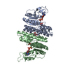
- PDB-7sce: Ternary complex of fixed-arm Trx-3ost5 (I299E) with 8mer-2 octasa... -
+ Open data

- Basic information
| Entry | Database: PDB / ID: 7sce | |||||||||||||||||||||
|---|---|---|---|---|---|---|---|---|---|---|---|---|---|---|---|---|---|---|---|---|---|---|
| Title | Ternary complex of fixed-arm Trx-3ost5 (I299E) with 8mer-2 octasaccharide substrate and co-factor product PAP | |||||||||||||||||||||
 Components Components | Thioredoxin 1,Heparan sulfate glucosamine 3-O-sulfotransferase 5 | |||||||||||||||||||||
 Keywords Keywords | TRANSFERASE/SUBSTRATE / sulfotransferase / heparan sulfate / oligosaccharide / complex / TRANSFERASE / TRANSFERASE-SUBSTRATE complex | |||||||||||||||||||||
| Function / homology |  Function and homology information Function and homology informationprotein sulfation / [heparan sulfate]-glucosamine 3-sulfotransferase 1 / [heparan sulfate]-glucosamine 3-sulfotransferase activity / regulation of viral entry into host cell / 3'-phosphoadenosine 5'-phosphosulfate binding / negative regulation of coagulation / HS-GAG biosynthesis / heparan sulfate proteoglycan biosynthetic process / DNA polymerase processivity factor activity / protein-disulfide reductase activity ...protein sulfation / [heparan sulfate]-glucosamine 3-sulfotransferase 1 / [heparan sulfate]-glucosamine 3-sulfotransferase activity / regulation of viral entry into host cell / 3'-phosphoadenosine 5'-phosphosulfate binding / negative regulation of coagulation / HS-GAG biosynthesis / heparan sulfate proteoglycan biosynthetic process / DNA polymerase processivity factor activity / protein-disulfide reductase activity / cell redox homeostasis / Golgi membrane / membrane / cytosol / cytoplasm Similarity search - Function | |||||||||||||||||||||
| Biological species |   Homo sapiens (human) Homo sapiens (human) | |||||||||||||||||||||
| Method |  X-RAY DIFFRACTION / SYNCHROTRON / MOLECULAR REPLACEMENT / Resolution: 2.75 Å X-RAY DIFFRACTION / SYNCHROTRON / MOLECULAR REPLACEMENT / Resolution: 2.75 Å | |||||||||||||||||||||
 Authors Authors | Wander, R. / Kaminski, A.M. / Krahn, J.M. / Liu, J. / Pedersen, L.C. | |||||||||||||||||||||
| Funding support |  United States, 6items United States, 6items
| |||||||||||||||||||||
 Citation Citation | Journal: Acs Catalysis / Year: 2021 Title: Structural and Substrate Specificity Analysis of 3-O-Sulfotransferase Isoform 5 to Synthesize Heparan Sulfate Authors: Wander, R. / Kaminski, A.M. / Wang, Z. / Stancanelli, E. / Xu, Y. / Pagadala, V. / Li, J. / Krahn, J.M. / Pham, T.Q. / Liu, J. / Pedersen, L.C. | |||||||||||||||||||||
| History |
|
- Structure visualization
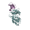
| Structure viewer | Molecule: MolmilJmol/JSmol |
|---|
- Downloads & links
-Download
| PDBx/mmCIF format | 7sce.cif.gz | 187 KB | Display | PDBx/mmCIF format |
|---|---|---|---|---|
| PDB format | pdb7sce.ent.gz | 127.8 KB | Display | PDB format |
| PDBx/mmJSON format | 7sce.json.gz | Tree view | PDBx/mmJSON format | |
| Others |  Other downloads Other downloads |
-Validation report
| Arichive directory | https://data.pdbj.org/pub/pdb/validation_reports/sc/7sceftp://data.pdbj.org/pub/pdb/validation_reports/sc/7sce | HTTPS FTP |
|---|
-Related structure data
| Related structure data |  7scdC  3bd9S S: Starting model for refinement C: citing same article ( |
|---|---|
| Similar structure data |


-Links
 PDBj
PDBj


- Assembly
| Deposited unit | 
| ||||||||||||
|---|---|---|---|---|---|---|---|---|---|---|---|---|---|
| 1 |
| ||||||||||||
| Unit cell |
|
-Components
| #1: Protein | Mass: 42821.449 Da / Num. of mol.: 1 / Mutation: I299E Source method: isolated from a genetically manipulated source Source: (gene. exp.) Homo sapiens (human)Strain: K12 Gene: trxA, fipA, tsnC, b3781, JW5856, HS3ST5, 3OST5, HS3OST5 Plasmid: pET32bX / Production host: References: UniProt: P0AA25, UniProt: Q8IZT8, [heparan sulfate]-glucosamine 3-sulfotransferase 1 |
|---|---|
| #2: Polysaccharide | 2-deoxy-6-O-sulfo-2-(sulfoamino)-alpha-D-glucopyranose-(1-4)-beta-D-glucopyranuronic acid-(1-4)-2- ...2-deoxy-6-O-sulfo-2-(sulfoamino)-alpha-D-glucopyranose-(1-4)-beta-D-glucopyranuronic acid-(1-4)-2-deoxy-6-O-sulfo-2-(sulfoamino)-alpha-D-glucopyranose-(1-4)-beta-D-glucopyranuronic acid-(1-4)-2-deoxy-6-O-sulfo-2-(sulfoamino)-alpha-D-glucopyranose-(1-4)-2-O-sulfo-alpha-L-idopyranuronic acid Type: oligosaccharide / Mass: 1590.299 Da / Num. of mol.: 1 / Source method: obtained synthetically |
| #3: Chemical | ChemComp-A3P / 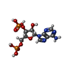  Type: RNA linking / Mass: 427.201 Da / Num. of mol.: 1 / Source method: obtained synthetically / Formula: C10H15N5O10P2 / Feature type: SUBJECT OF INVESTIGATION Type: RNA linking / Mass: 427.201 Da / Num. of mol.: 1 / Source method: obtained synthetically / Formula: C10H15N5O10P2 / Feature type: SUBJECT OF INVESTIGATION |
| #4: Water | ChemComp-HOH /  Mass: 18.015 Da / Num. of mol.: 53 / Source method: isolated from a natural source / Formula: H2O Mass: 18.015 Da / Num. of mol.: 53 / Source method: isolated from a natural source / Formula: H2O |
| Has ligand of interest | Y |
| Has protein modification | Y |
-Experimental details
-Experiment
| Experiment | Method: X-RAY DIFFRACTION / Number of used crystals: 1 |
|---|
- Sample preparation
| Crystal | Density Matthews: 2.9 Å3/Da / Density % sol: 57.6 % |
|---|---|
| Crystal grow | Temperature: 295 K / Method: vapor diffusion, sitting drop / pH: 4.6 Details: 85mM sodium acetate pH 4.6, 0.17M ammonium acetate, 25.5% (w/v) PEG4000, 15% (v/v) ethylene glycol |
-Data collection
| Diffraction | Mean temperature: 93 K / Serial crystal experiment: N |
|---|---|
| Diffraction source | Source: SYNCHROTRON / Site: APS / Beamline: 22-ID / Wavelength: 1 Å |
| Detector | Type: DECTRIS EIGER X 16M / Detector: PIXEL / Date: Jun 10, 2021 |
| Radiation | Monochromator: DOUBLE CRYSTAL SI(111) / Protocol: SINGLE WAVELENGTH / Monochromatic (M) / Laue (L): M / Scattering type: x-ray |
| Radiation wavelength | Wavelength: 1 Å / Relative weight: 1 |
| Reflection | Resolution: 2.75→50 Å / Num. obs: 13239 / % possible obs: 99.1 % / Redundancy: 4.9 % / Biso Wilson estimate: 31.65 Å2 / CC1/2: 0.992 / CC star: 0.998 / Rsym value: 0.163 / Net I/σ(I): 7.46 |
| Reflection shell | Resolution: 2.75→2.8 Å / Mean I/σ(I) obs: 1.54 / Num. unique obs: 642 / CC1/2: 0.661 / CC star: 0.892 / Rsym value: 0.671 |
- Processing
| Software |
| |||||||||||||||||||||||||||||||||||||||||||||||||||||||||||||||||||||||||||||||||||||||||||||||||||||||||||||||||||||||||||||
|---|---|---|---|---|---|---|---|---|---|---|---|---|---|---|---|---|---|---|---|---|---|---|---|---|---|---|---|---|---|---|---|---|---|---|---|---|---|---|---|---|---|---|---|---|---|---|---|---|---|---|---|---|---|---|---|---|---|---|---|---|---|---|---|---|---|---|---|---|---|---|---|---|---|---|---|---|---|---|---|---|---|---|---|---|---|---|---|---|---|---|---|---|---|---|---|---|---|---|---|---|---|---|---|---|---|---|---|---|---|---|---|---|---|---|---|---|---|---|---|---|---|---|---|---|---|---|
| Refinement | Method to determine structure: MOLECULAR REPLACEMENT Starting model: 3BD9 Resolution: 2.75→40 Å / SU ML: 0.3285 / Cross valid method: FREE R-VALUE / σ(F): 1.35 / Phase error: 21.8839 Stereochemistry target values: GeoStd + Monomer Library + CDL v1.2
| |||||||||||||||||||||||||||||||||||||||||||||||||||||||||||||||||||||||||||||||||||||||||||||||||||||||||||||||||||||||||||||
| Solvent computation | Shrinkage radii: 0.9 Å / VDW probe radii: 1.11 Å / Solvent model: FLAT BULK SOLVENT MODEL | |||||||||||||||||||||||||||||||||||||||||||||||||||||||||||||||||||||||||||||||||||||||||||||||||||||||||||||||||||||||||||||
| Displacement parameters | Biso mean: 31.26 Å2 | |||||||||||||||||||||||||||||||||||||||||||||||||||||||||||||||||||||||||||||||||||||||||||||||||||||||||||||||||||||||||||||
| Refinement step | Cycle: LAST / Resolution: 2.75→40 Å
| |||||||||||||||||||||||||||||||||||||||||||||||||||||||||||||||||||||||||||||||||||||||||||||||||||||||||||||||||||||||||||||
| Refine LS restraints |
| |||||||||||||||||||||||||||||||||||||||||||||||||||||||||||||||||||||||||||||||||||||||||||||||||||||||||||||||||||||||||||||
| LS refinement shell |
| |||||||||||||||||||||||||||||||||||||||||||||||||||||||||||||||||||||||||||||||||||||||||||||||||||||||||||||||||||||||||||||
| Refinement TLS params. | Method: refined / Refine-ID: X-RAY DIFFRACTION
| |||||||||||||||||||||||||||||||||||||||||||||||||||||||||||||||||||||||||||||||||||||||||||||||||||||||||||||||||||||||||||||
| Refinement TLS group | Refine-ID: X-RAY DIFFRACTION
|
