 Movie
Movie Controller

[English] 日本語
 Yorodumi
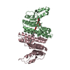
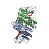
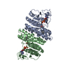
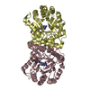
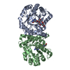
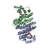
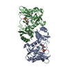
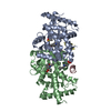
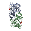
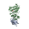
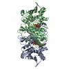
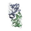
- PDB-7lnt: Ternary complex of the Isopentenyl Phosphate Kinase from Candidat... -
+ Open data

- Basic information
| Entry | Database: PDB / ID: 7lnt | ||||||
|---|---|---|---|---|---|---|---|
| Title | Ternary complex of the Isopentenyl Phosphate Kinase from Candidatus methanomethylophilus alvus bound to benzyl monophosphate and ATP | ||||||
 Components Components | Isopentenyl phosphate kinase | ||||||
 Keywords Keywords | TRANSFERASE / Phosphotransferase ATP Biocatalysis Isoprenoids Enzyme Promiscuity | ||||||
| Function / homology |  Function and homology information Function and homology informationisopentenyl phosphate kinase / isopentenyl phosphate kinase activity / terpenoid biosynthetic process / kinase activity / ATP binding / cytosol Similarity search - Function | ||||||
| Biological species |  Candidatus Methanomethylophilus alvus (archaea) Candidatus Methanomethylophilus alvus (archaea) | ||||||
| Method |  X-RAY DIFFRACTION / MOLECULAR REPLACEMENT / Resolution: 2.35 Å X-RAY DIFFRACTION / MOLECULAR REPLACEMENT / Resolution: 2.35 Å | ||||||
 Authors Authors | Thomas, L.M. / Singh, S. / Scull, E.M. / Bourne, C.R. | ||||||
| Funding support |  United States, 1items United States, 1items
| ||||||
 Citation Citation | Journal: Acs Chem.Biol. / Year: 2022 Title: Molecular Basis for the Substrate Promiscuity of Isopentenyl Phosphate Kinase from Candidatus methanomethylophilus alvus . Authors: Johnson, B.P. / Kumar, V. / Scull, E.M. / Thomas, L.M. / Bourne, C.R. / Singh, S. | ||||||
| History |
|
- Structure visualization
| Structure viewer | Molecule: MolmilJmol/JSmol |
|---|
- Downloads & links
-Download
| PDBx/mmCIF format | 7lnt.cif.gz | 142.1 KB | Display | PDBx/mmCIF format |
|---|---|---|---|---|
| PDB format | pdb7lnt.ent.gz | 88.3 KB | Display | PDB format |
| PDBx/mmJSON format | 7lnt.json.gz | Tree view | PDBx/mmJSON format | |
| Others |  Other downloads Other downloads |
-Validation report
| Arichive directory | https://data.pdbj.org/pub/pdb/validation_reports/ln/7lntftp://data.pdbj.org/pub/pdb/validation_reports/ln/7lnt | HTTPS FTP |
|---|
-Related structure data


-Links
 PDBj
PDBj- Assembly
| Deposited unit | 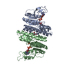
| ||||||||||||
|---|---|---|---|---|---|---|---|---|---|---|---|---|---|
| 1 |
| ||||||||||||
| Unit cell |
|
-Components
| #1: Protein | Mass: 30533.225 Da / Num. of mol.: 2 Source method: isolated from a genetically manipulated source Source: (gene. exp.) Candidatus Methanomethylophilus alvus (archaea)Gene: BKD89_07040 / Production host:  References: UniProt: A0A3G3II74, isopentenyl phosphate kinase #2: Chemical |   Mass: 92.094 Da / Num. of mol.: 2 / Source method: obtained synthetically / Formula: C3H8O3 Mass: 92.094 Da / Num. of mol.: 2 / Source method: obtained synthetically / Formula: C3H8O3#3: Chemical | 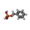  Mass: 188.118 Da / Num. of mol.: 2 / Source method: obtained synthetically / Formula: C7H9O4P / Feature type: SUBJECT OF INVESTIGATION Mass: 188.118 Da / Num. of mol.: 2 / Source method: obtained synthetically / Formula: C7H9O4P / Feature type: SUBJECT OF INVESTIGATION#4: Chemical |   Mass: 427.201 Da / Num. of mol.: 2 / Source method: obtained synthetically / Formula: C10H15N5O10P2 / Comment: ADP, energy-carrying molecule*YM Mass: 427.201 Da / Num. of mol.: 2 / Source method: obtained synthetically / Formula: C10H15N5O10P2 / Comment: ADP, energy-carrying molecule*YM#5: Water | ChemComp-HOH / |  Mass: 18.015 Da / Num. of mol.: 209 / Source method: isolated from a natural source / Formula: H2O Mass: 18.015 Da / Num. of mol.: 209 / Source method: isolated from a natural source / Formula: H2OHas ligand of interest | Y | |
|---|
-Experimental details
-Experiment
| Experiment | Method: X-RAY DIFFRACTION / Number of used crystals: 1 |
|---|
- Sample preparation
| Crystal | Density Matthews: 2.13 Å3/Da / Density % sol: 42.23 % |
|---|---|
| Crystal grow | Temperature: 295 K / Method: vapor diffusion / Details: 0.1 M Bis-Tris pH 5.5 15% PEG 10000 |
-Data collection
| Diffraction | Mean temperature: 100 K / Serial crystal experiment: N |
|---|---|
| Diffraction source | Source: ROTATING ANODE / Type: RIGAKU MICROMAX-007 HF / Wavelength: 1.54178 Å |
| Detector | Type: DECTRIS PILATUS 200K / Detector: PIXEL / Date: Sep 22, 2016 / Details: Osmic Veri Max Mirror |
| Radiation | Protocol: SINGLE WAVELENGTH / Monochromatic (M) / Laue (L): M / Scattering type: x-ray |
| Radiation wavelength | Wavelength: 1.54178 Å / Relative weight: 1 |
| Reflection | Resolution: 2.35→30 Å / Num. obs: 22277 / % possible obs: 98.9 % / Redundancy: 4.5 % / Biso Wilson estimate: 28.17 Å2 / Rpim(I) all: 0.048 / Rrim(I) all: 0.109 / Χ2: 1.037 / Net I/σ(I): 16.9 |
| Reflection shell | Resolution: 2.35→2.39 Å / Mean I/σ(I) obs: 2.89 / Num. unique obs: 999 / CC1/2: 0.874 / Rpim(I) all: 0.221 / Rrim(I) all: 0.412 / Χ2: 0.712 / % possible all: 89.5 |
- Processing
| Software |
| |||||||||||||||||||||||||||||||||||||||||||||||||||||||||||||||
|---|---|---|---|---|---|---|---|---|---|---|---|---|---|---|---|---|---|---|---|---|---|---|---|---|---|---|---|---|---|---|---|---|---|---|---|---|---|---|---|---|---|---|---|---|---|---|---|---|---|---|---|---|---|---|---|---|---|---|---|---|---|---|---|---|
| Refinement | Method to determine structure: MOLECULAR REPLACEMENT Starting model: CMA apo model Resolution: 2.35→29.45 Å / SU ML: 0.2472 / Cross valid method: FREE R-VALUE / σ(F): 1.36 / Phase error: 22.529 Stereochemistry target values: GeoStd + Monomer Library + CDL v1.2
| |||||||||||||||||||||||||||||||||||||||||||||||||||||||||||||||
| Solvent computation | Shrinkage radii: 0.9 Å / VDW probe radii: 1.11 Å / Solvent model: FLAT BULK SOLVENT MODEL | |||||||||||||||||||||||||||||||||||||||||||||||||||||||||||||||
| Displacement parameters | Biso mean: 33.84 Å2 | |||||||||||||||||||||||||||||||||||||||||||||||||||||||||||||||
| Refinement step | Cycle: LAST / Resolution: 2.35→29.45 Å
| |||||||||||||||||||||||||||||||||||||||||||||||||||||||||||||||
| Refine LS restraints |
| |||||||||||||||||||||||||||||||||||||||||||||||||||||||||||||||
| LS refinement shell |
|
