 ムービー
ムービー コントローラー


+ データを開く

- 基本情報
基本情報
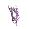
| 登録情報 | データベース: PDB / ID: 7s7s | ||||||
|---|---|---|---|---|---|---|---|
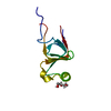
| タイトル | Crystal structure of hydrophobin SC16, P21212 | ||||||
 要素 要素 | Hydrophobin | ||||||
 キーワード キーワード | STRUCTURAL PROTEIN / hydrophobin / self-assembly / surface modifier | ||||||
| 機能・相同性 | Fungal hydrophobin / Hydrophobin, conserved site / Fungal hydrophobins signature. / Hydrophobin / Hydrophobins / structural constituent of cell wall / fungal-type cell wall / extracellular region / Class I hydrophobin SC16 機能・相同性情報 機能・相同性情報 | ||||||
| 生物種 |  Schizophyllum commune (スエヒロタケ) Schizophyllum commune (スエヒロタケ) | ||||||
| 手法 |  X線回折 / 分子置換 / 解像度: 2.2 Å X線回折 / 分子置換 / 解像度: 2.2 Å | ||||||
 データ登録者 データ登録者 | Vergunst, K.L. / Langelaan, D.N. | ||||||
| 資金援助 |  カナダ, 1件 カナダ, 1件
| ||||||
 引用 引用 | ジャーナル: Sci Rep / 年: 2022 タイトル: The N-terminal tail of the hydrophobin SC16 is not required for rodlet formation. 著者: Vergunst, K.L. / Langelaan, D.N. #1: ジャーナル: Acta Crystallogr.,Sect.D / 年: 2012 タイトル: Towards automated crystallographic structure refinement with phenix.refine. 著者: Afonine, P.V. / Grosse-Kunstleve, R.W. / Echols, N. / Headd, J.J. / Moriarty, N.W. / Mustyakimov, M. / Terwilliger, T.C. / Urzhumtsev, A. / Zwart, P.H. / Adams, P.D. #2: ジャーナル: Acta Crystallogr D Struct Biol / 年: 2019 タイトル: Macromolecular structure determination using X-rays, neutrons and electrons: recent developments in Phenix. 著者: Dorothee Liebschner / Pavel V Afonine / Matthew L Baker / Gábor Bunkóczi / Vincent B Chen / Tristan I Croll / Bradley Hintze / Li Wei Hung / Swati Jain / Airlie J McCoy / Nigel W Moriarty / ...著者: Dorothee Liebschner / Pavel V Afonine / Matthew L Baker / Gábor Bunkóczi / Vincent B Chen / Tristan I Croll / Bradley Hintze / Li Wei Hung / Swati Jain / Airlie J McCoy / Nigel W Moriarty / Robert D Oeffner / Billy K Poon / Michael G Prisant / Randy J Read / Jane S Richardson / David C Richardson / Massimo D Sammito / Oleg V Sobolev / Duncan H Stockwell / Thomas C Terwilliger / Alexandre G Urzhumtsev / Lizbeth L Videau / Christopher J Williams / Paul D Adams /    要旨: Diffraction (X-ray, neutron and electron) and electron cryo-microscopy are powerful methods to determine three-dimensional macromolecular structures, which are required to understand biological ...Diffraction (X-ray, neutron and electron) and electron cryo-microscopy are powerful methods to determine three-dimensional macromolecular structures, which are required to understand biological processes and to develop new therapeutics against diseases. The overall structure-solution workflow is similar for these techniques, but nuances exist because the properties of the reduced experimental data are different. Software tools for structure determination should therefore be tailored for each method. Phenix is a comprehensive software package for macromolecular structure determination that handles data from any of these techniques. Tasks performed with Phenix include data-quality assessment, map improvement, model building, the validation/rebuilding/refinement cycle and deposition. Each tool caters to the type of experimental data. The design of Phenix emphasizes the automation of procedures, where possible, to minimize repetitive and time-consuming manual tasks, while default parameters are chosen to encourage best practice. A graphical user interface provides access to many command-line features of Phenix and streamlines the transition between programs, project tracking and re-running of previous tasks. | ||||||
| 履歴 |
|
- 構造の表示
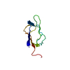
構造の表示
| 構造ビューア | 分子: MolmilJmol/JSmol |
|---|
- ダウンロードとリンク
-ダウンロード
| PDBx/mmCIF形式 | 7s7s.cif.gz | 50.3 KB | 表示 | PDBx/mmCIF形式 |
|---|---|---|---|---|
| PDB形式 | pdb7s7s.ent.gz | 29.2 KB | 表示 | PDB形式 |
| PDBx/mmJSON形式 | 7s7s.json.gz | ツリー表示 | PDBx/mmJSON形式 | |
| その他 |  その他のダウンロード その他のダウンロード |
-検証レポート
| 文書・要旨 | 7s7s_validation.pdf.gz | 424.3 KB | 表示 | wwPDB検証レポート |
|---|---|---|---|---|
| 文書・詳細版 | 7s7s_full_validation.pdf.gz | 424.4 KB | 表示 | |
| XML形式データ | 7s7s_validation.xml.gz | 6.1 KB | 表示 | |
| CIF形式データ | 7s7s_validation.cif.gz | 7.6 KB | 表示 | |
| アーカイブディレクトリ | https://data.pdbj.org/pub/pdb/validation_reports/s7/7s7sftp://data.pdbj.org/pub/pdb/validation_reports/s7/7s7s | HTTPS FTP |
-関連構造データ
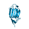
| 関連構造データ | 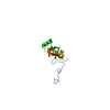 2nbhS S: 精密化の開始モデル |
|---|---|
| 類似構造データ |


-リンク
 PDBj
PDBj- 集合体
集合体
| 登録構造単位 | 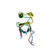
| ||||||||||||
|---|---|---|---|---|---|---|---|---|---|---|---|---|---|
| 1 |
| ||||||||||||
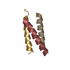
| 単位格子 |
|
-要素
| #1: タンパク質 | 分子量: 10098.559 Da / 分子数: 1 / 由来タイプ: 組換発現 詳細: A naturally occurring point mutant that differs from UniProt (insertion of Ser30 and Lys31). 由来: (組換発現) Schizophyllum commune (スエヒロタケ)遺伝子: HYD1, SCHCODRAFT_58269 / 発現宿主:  |
|---|---|
| #2: 水 | ChemComp-HOH /  分子量: 18.015 Da / 分子数: 63 / 由来タイプ: 天然 / 式: H2O 分子量: 18.015 Da / 分子数: 63 / 由来タイプ: 天然 / 式: H2O |
| Has protein modification | Y |
-実験情報
-実験
| 実験 | 手法: X線回折 / 使用した結晶の数: 1 |
|---|
- 試料調製
試料調製
| 結晶 | マシュー密度: 1.79 Å3/Da / 溶媒含有率: 37.83 % / 解説: Large clusters of plates |
|---|---|
| 結晶化 | 温度: 293 K / 手法: 蒸気拡散法, ハンギングドロップ法 / pH: 3.5 / 詳細: 0.1 M Citric Acid pH 3.5 29% PEG 3350 |
-データ収集
| 回折 | 平均測定温度: 100 K / Serial crystal experiment: N |
|---|---|
| 放射光源 | 由来: SEALED TUBE / タイプ: BRUKER D8 QUEST / 波長: 1.5406 Å |
| 検出器 | タイプ: Bruker PHOTON II / 検出器: PIXEL / 日付: 2020年3月20日 |
| 放射 | プロトコル: SINGLE WAVELENGTH / 単色(M)・ラウエ(L): M / 散乱光タイプ: x-ray |
| 放射波長 | 波長: 1.5406 Å / 相対比: 1 |
| 反射 | 解像度: 2.2→21.881 Å / Num. obs: 4374 / % possible obs: 100 % / 冗長度: 58.3 % / Biso Wilson estimate: 20.03 Å2 / CC1/2: 0.999 / Rmerge(I) obs: 0.196 / Rpim(I) all: 0.026 / Rrim(I) all: 0.198 / Net I/σ(I): 21.4 |
| 反射 シェル | 解像度: 2.2→2.28 Å / Rmerge(I) obs: 0.783 / Mean I/σ(I) obs: 6.3 / Num. unique obs: 422 / CC1/2: 0.98 / Rpim(I) all: 0.101 / Rrim(I) all: 0.79 / % possible all: 100 |
- 解析
解析
| ソフトウェア |
| ||||||||||||||||||||||||
|---|---|---|---|---|---|---|---|---|---|---|---|---|---|---|---|---|---|---|---|---|---|---|---|---|---|
| 精密化 | 構造決定の手法: 分子置換 開始モデル: 2NBH 解像度: 2.2→21.21 Å / SU ML: 0.1719 / 交差検証法: FREE R-VALUE / σ(F): 0 / 位相誤差: 24.8295 立体化学のターゲット値: GeoStd + Monomer Library + CDL v1.2
| ||||||||||||||||||||||||
| 溶媒の処理 | 減衰半径: 0.9 Å / VDWプローブ半径: 1.11 Å / 溶媒モデル: FLAT BULK SOLVENT MODEL | ||||||||||||||||||||||||
| 原子変位パラメータ | Biso mean: 26.21 Å2 | ||||||||||||||||||||||||
| 精密化ステップ | サイクル: LAST / 解像度: 2.2→21.21 Å
| ||||||||||||||||||||||||
| 拘束条件 |
| ||||||||||||||||||||||||
| LS精密化 シェル | 解像度: 2.2→2.279 Å
|
