 Movie
Movie Controller

[English] 日本語
 Yorodumi
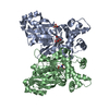
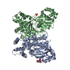
- PDB-7s2n: Crystal structure of the F337L mutation of Trypanosoma cruzi gluc... -
+ Open data

- Basic information
| Entry | Database: PDB / ID: 7s2n | ||||||
|---|---|---|---|---|---|---|---|
| Title | Crystal structure of the F337L mutation of Trypanosoma cruzi glucokinase in the apo form (open conformation) | ||||||
 Components Components | Glucokinase 1 | ||||||
 Keywords Keywords | TRANSFERASE / Trypanosoma cruzi / Chagas disease | ||||||
| Function / homology | Glucokinase / Glucokinase / glucokinase / glucokinase activity / D-glucose binding / glycolytic process / ATPase, nucleotide binding domain / ATP binding / Glucokinase 1, putative Function and homology information Function and homology information | ||||||
| Biological species |  | ||||||
| Method |  X-RAY DIFFRACTION / SYNCHROTRON / MOLECULAR REPLACEMENT / Resolution: 1.75 Å X-RAY DIFFRACTION / SYNCHROTRON / MOLECULAR REPLACEMENT / Resolution: 1.75 Å | ||||||
 Authors Authors | Carey, S.M. / Nettles, R.B. / Daneshian, L. / Chruszcz, M. / D'Antonio, E.L. | ||||||
| Funding support |  United States, 1items United States, 1items
| ||||||
 Citation Citation | Journal: To Be Published Title: Crystal structure of the F337L mutation of Trypanosoma cruzi glucokinase in the apo form (open conformation) Authors: Carey, S.M. / Nettles, R.B. / Daneshian, L. / Chruszcz, M. / D'Antonio, E.L. | ||||||
| History |
|
- Structure visualization
| Structure viewer | Molecule: MolmilJmol/JSmol |
|---|
- Downloads & links
-Download
| PDBx/mmCIF format | 7s2n.cif.gz | 310.8 KB | Display | PDBx/mmCIF format |
|---|---|---|---|---|
| PDB format | pdb7s2n.ent.gz | 250.8 KB | Display | PDB format |
| PDBx/mmJSON format | 7s2n.json.gz | Tree view | PDBx/mmJSON format | |
| Others |  Other downloads Other downloads |
-Validation report
| Arichive directory | https://data.pdbj.org/pub/pdb/validation_reports/s2/7s2nftp://data.pdbj.org/pub/pdb/validation_reports/s2/7s2n | HTTPS FTP |
|---|
-Related structure data
| Related structure data | 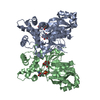 2q2rS S: Starting model for refinement |
|---|---|
| Similar structure data |


-Links
 PDBj
PDBj- Assembly
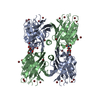
| Deposited unit | 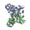
| ||||||||||||||||||
|---|---|---|---|---|---|---|---|---|---|---|---|---|---|---|---|---|---|---|---|
| 1 |
| ||||||||||||||||||
| Unit cell |
| ||||||||||||||||||
| Noncrystallographic symmetry (NCS) | NCS domain:
NCS domain segments: Component-ID: _ / Ens-ID: 1 / Beg auth comp-ID: ALA / Beg label comp-ID: ALA / End auth comp-ID: ASP / End label comp-ID: ASP / Refine code: _ / Auth seq-ID: 0 - 365 / Label seq-ID: 14 - 379
|
-Components
| #1: Protein | Mass: 42192.633 Da / Num. of mol.: 2 / Mutation: F337L Source method: isolated from a genetically manipulated source Source: (gene. exp.) Strain: CL Brener / Gene: Tc00.1047053510187.100 / Production host:  #2: Water | ChemComp-HOH / |  Mass: 18.015 Da / Num. of mol.: 729 / Source method: isolated from a natural source / Formula: H2O Mass: 18.015 Da / Num. of mol.: 729 / Source method: isolated from a natural source / Formula: H2O |
|---|
-Experimental details
-Experiment
| Experiment | Method: X-RAY DIFFRACTION / Number of used crystals: 1 |
|---|
- Sample preparation
| Crystal | Density Matthews: 2.41 Å3/Da / Density % sol: 49.02 % |
|---|---|
| Crystal grow | Temperature: 295 K / Method: vapor diffusion, sitting drop / pH: 7.5 Details: 1.0 uL of 7.4 mg/mL TcGlcK(F337L) in buffered solution [50 mM HEPES (pH 7.5), 0.2 M imidazole, 2 mM magnesium chloride] + 1.0 uL of precipitant solution [14% (w/v) PEG 3350, 0.1 M sodium ...Details: 1.0 uL of 7.4 mg/mL TcGlcK(F337L) in buffered solution [50 mM HEPES (pH 7.5), 0.2 M imidazole, 2 mM magnesium chloride] + 1.0 uL of precipitant solution [14% (w/v) PEG 3350, 0.1 M sodium citrate tribasic] was equilibrated against 85 uL of the precipitant solution using a 96-well sitting-drop plate (Innovadyne) |
-Data collection
| Diffraction | Mean temperature: 100 K / Serial crystal experiment: N |
|---|---|
| Diffraction source | Source: SYNCHROTRON / Site: APS / Beamline: 14-ID-B / Wavelength: 0.9792 Å |
| Detector | Type: ADSC QUANTUM 315r / Detector: CCD / Date: Jun 27, 2014 |
| Radiation | Protocol: SINGLE WAVELENGTH / Monochromatic (M) / Laue (L): M / Scattering type: x-ray |
| Radiation wavelength | Wavelength: 0.9792 Å / Relative weight: 1 |
| Reflection | Resolution: 1.75→40 Å / Num. obs: 80560 / % possible obs: 99.9 % / Observed criterion σ(I): -3 / Redundancy: 3.8 % / Rpim(I) all: 0.031 / Rrim(I) all: 0.059 / Net I/σ(I): 32.7 |
| Reflection shell | Resolution: 1.75→1.78 Å / Redundancy: 3.7 % / Mean I/σ(I) obs: 2 / Num. unique obs: 4061 / CC1/2: 0.688 / Rpim(I) all: 0.4 / Rrim(I) all: 0.774 / % possible all: 100 |
- Processing
| Software |
| |||||||||||||||||||||||||||||||||||||||||||||||||||||||||||||||||||||||||||||||||||||||||||||||||||||||||||||||||||||||||||||
|---|---|---|---|---|---|---|---|---|---|---|---|---|---|---|---|---|---|---|---|---|---|---|---|---|---|---|---|---|---|---|---|---|---|---|---|---|---|---|---|---|---|---|---|---|---|---|---|---|---|---|---|---|---|---|---|---|---|---|---|---|---|---|---|---|---|---|---|---|---|---|---|---|---|---|---|---|---|---|---|---|---|---|---|---|---|---|---|---|---|---|---|---|---|---|---|---|---|---|---|---|---|---|---|---|---|---|---|---|---|---|---|---|---|---|---|---|---|---|---|---|---|---|---|---|---|---|
| Refinement | Method to determine structure: MOLECULAR REPLACEMENT Starting model: 2Q2R Resolution: 1.75→39.69 Å / Cor.coef. Fo:Fc: 0.972 / Cor.coef. Fo:Fc free: 0.959 / SU B: 4.47 / SU ML: 0.072 / Cross valid method: THROUGHOUT / σ(F): 0 / ESU R: 0.102 / ESU R Free: 0.099 / Stereochemistry target values: MAXIMUM LIKELIHOOD Details: HYDROGENS HAVE BEEN ADDED IN THE RIDING POSITIONS U VALUES : WITH TLS ADDED
| |||||||||||||||||||||||||||||||||||||||||||||||||||||||||||||||||||||||||||||||||||||||||||||||||||||||||||||||||||||||||||||
| Solvent computation | Ion probe radii: 0.8 Å / Shrinkage radii: 0.8 Å / VDW probe radii: 1.2 Å / Solvent model: BABINET MODEL WITH MASK | |||||||||||||||||||||||||||||||||||||||||||||||||||||||||||||||||||||||||||||||||||||||||||||||||||||||||||||||||||||||||||||
| Displacement parameters | Biso max: 99.3 Å2 / Biso mean: 34.491 Å2 / Biso min: 18.92 Å2
| |||||||||||||||||||||||||||||||||||||||||||||||||||||||||||||||||||||||||||||||||||||||||||||||||||||||||||||||||||||||||||||
| Refinement step | Cycle: final / Resolution: 1.75→39.69 Å
| |||||||||||||||||||||||||||||||||||||||||||||||||||||||||||||||||||||||||||||||||||||||||||||||||||||||||||||||||||||||||||||
| Refine LS restraints |
| |||||||||||||||||||||||||||||||||||||||||||||||||||||||||||||||||||||||||||||||||||||||||||||||||||||||||||||||||||||||||||||
| Refine LS restraints NCS | Ens-ID: 1 / Number: 11256 / Refine-ID: X-RAY DIFFRACTION / Type: interatomic distance / Rms dev position: 0.11 Å / Weight position: 0.05
| |||||||||||||||||||||||||||||||||||||||||||||||||||||||||||||||||||||||||||||||||||||||||||||||||||||||||||||||||||||||||||||
| LS refinement shell | Resolution: 1.75→1.794 Å / Rfactor Rfree error: 0
| |||||||||||||||||||||||||||||||||||||||||||||||||||||||||||||||||||||||||||||||||||||||||||||||||||||||||||||||||||||||||||||
| Refinement TLS params. | Method: refined / Refine-ID: X-RAY DIFFRACTION
| |||||||||||||||||||||||||||||||||||||||||||||||||||||||||||||||||||||||||||||||||||||||||||||||||||||||||||||||||||||||||||||
| Refinement TLS group |
|
