 Movie
Movie Controller

[English] 日本語
 Yorodumi
- PDB-7rk1: Crystal structure of the human astrovirus serotype 8 capsid spike... -
+ Open data

- Basic information
| Entry | Database: PDB / ID: 7rk1 | ||||||
|---|---|---|---|---|---|---|---|
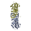
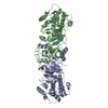
| Title | Crystal structure of the human astrovirus serotype 8 capsid spike in complex with scFv 3E8, an astrovirus-neutralizing antibody, at 2.05-A resolution | ||||||
 Components Components |
| ||||||
 Keywords Keywords | VIRAL PROTEIN / capsid protein / icosahedral virus / single chain variable fragment | ||||||
| Function / homology |  Function and homology information Function and homology informationT=3 icosahedral viral capsid / clathrin-dependent endocytosis of virus by host cell / host extracellular space / identical protein binding Similarity search - Function | ||||||
| Biological species |  Human astrovirus-8 Human astrovirus-8 | ||||||
| Method |  X-RAY DIFFRACTION / SYNCHROTRON / MOLECULAR REPLACEMENT / Resolution: 2.05 Å X-RAY DIFFRACTION / SYNCHROTRON / MOLECULAR REPLACEMENT / Resolution: 2.05 Å | ||||||
 Authors Authors | Meyer, L. / DuBois, R.M. | ||||||
| Funding support |  United States, 1items United States, 1items
| ||||||
 Citation Citation | Journal: J.Virol. / Year: 2022 Title: Structures of Two Human Astrovirus Capsid/Neutralizing Antibody Complexes Reveal Distinct Epitopes and Inhibition of Virus Attachment to Cells. Authors: Ricemeyer, L. / Aguilar-Hernandez, N. / Lopez, T. / Espinosa, R. / Lanning, S. / Mukherjee, S. / Cuellar, C. / Lopez, S. / Arias, C.F. / DuBois, R.M. | ||||||
| History |
|
- Structure visualization
| Structure viewer | Molecule: MolmilJmol/JSmol |
|---|
- Downloads & links
-Download
| PDBx/mmCIF format | 7rk1.cif.gz | 193.6 KB | Display | PDBx/mmCIF format |
|---|---|---|---|---|
| PDB format | pdb7rk1.ent.gz | 150.7 KB | Display | PDB format |
| PDBx/mmJSON format | 7rk1.json.gz | Tree view | PDBx/mmJSON format | |
| Others |  Other downloads Other downloads |
-Validation report
| Summary document | 7rk1_validation.pdf.gz | 454.4 KB | Display | wwPDB validaton report |
|---|---|---|---|---|
| Full document | 7rk1_full_validation.pdf.gz | 463.4 KB | Display | |
| Data in XML | 7rk1_validation.xml.gz | 34.2 KB | Display | |
| Data in CIF | 7rk1_validation.cif.gz | 49.1 KB | Display | |
| Arichive directory | https://data.pdbj.org/pub/pdb/validation_reports/rk/7rk1ftp://data.pdbj.org/pub/pdb/validation_reports/rk/7rk1 | HTTPS FTP |
-Related structure data
| Related structure data | 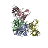 7rk2C 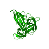 3qsqS S: Starting model for refinement C: citing same article ( |
|---|---|
| Similar structure data |


-Links
 PDBj
PDBj


- Assembly
| Deposited unit | 
| ||||||||
|---|---|---|---|---|---|---|---|---|---|
| 1 |
| ||||||||
| Unit cell |
|
-Components
| #1: Protein | Mass: 25886.105 Da / Num. of mol.: 2 Source method: isolated from a genetically manipulated source Source: (gene. exp.) Human astrovirus-8 / Gene: ORF2 / Production host:  #2: Antibody | Mass: 26878.584 Da / Num. of mol.: 2 Source method: isolated from a genetically manipulated source Source: (gene. exp.)  Cricetulus griseus (Chinese hamster) Cricetulus griseus (Chinese hamster)#3: Water | ChemComp-HOH / |  Mass: 18.015 Da / Num. of mol.: 335 / Source method: isolated from a natural source / Formula: H2O Mass: 18.015 Da / Num. of mol.: 335 / Source method: isolated from a natural source / Formula: H2OHas protein modification | Y | |
|---|
-Experimental details
-Experiment
| Experiment | Method: X-RAY DIFFRACTION / Number of used crystals: 1 |
|---|
- Sample preparation
| Crystal | Density Matthews: 2.18 Å3/Da / Density % sol: 43.6 % |
|---|---|
| Crystal grow | Temperature: 295.15 K / Method: vapor diffusion, hanging drop / Details: 0.2 M lithium citrate tribasic, 18 % PEG 3350 |
-Data collection
| Diffraction | Mean temperature: 100 K / Serial crystal experiment: N | ||||||||||||||||||||||||||||||
|---|---|---|---|---|---|---|---|---|---|---|---|---|---|---|---|---|---|---|---|---|---|---|---|---|---|---|---|---|---|---|---|
| Diffraction source | Source: SYNCHROTRON / Site: APS / Beamline: 23-ID-B / Wavelength: 1.033184 Å | ||||||||||||||||||||||||||||||
| Detector | Type: DECTRIS EIGER X 16M / Detector: PIXEL / Date: Mar 5, 2021 | ||||||||||||||||||||||||||||||
| Radiation | Monochromator: Si(111) / Protocol: SINGLE WAVELENGTH / Monochromatic (M) / Laue (L): M / Scattering type: x-ray | ||||||||||||||||||||||||||||||
| Radiation wavelength | Wavelength: 1.033184 Å / Relative weight: 1 | ||||||||||||||||||||||||||||||
| Reflection | Resolution: 2.05→80.43 Å / Num. obs: 55739 / % possible obs: 97.9 % / Redundancy: 6.1 % / Biso Wilson estimate: 25.28 Å2 / CC1/2: 0.992 / Rmerge(I) obs: 0.184 / Rpim(I) all: 0.081 / Rrim(I) all: 0.202 / Net I/σ(I): 7.2 / Num. measured all: 337857 / Scaling rejects: 795 | ||||||||||||||||||||||||||||||
| Reflection shell | Diffraction-ID: 1
|
- Processing
| Software |
| |||||||||||||||||||||||||||||||||||||||||||||||||||||||||||||||||||||||||||||||||||||||||||||||||||||||||
|---|---|---|---|---|---|---|---|---|---|---|---|---|---|---|---|---|---|---|---|---|---|---|---|---|---|---|---|---|---|---|---|---|---|---|---|---|---|---|---|---|---|---|---|---|---|---|---|---|---|---|---|---|---|---|---|---|---|---|---|---|---|---|---|---|---|---|---|---|---|---|---|---|---|---|---|---|---|---|---|---|---|---|---|---|---|---|---|---|---|---|---|---|---|---|---|---|---|---|---|---|---|---|---|---|---|---|
| Refinement | Method to determine structure: MOLECULAR REPLACEMENT Starting model: 3QSQ Resolution: 2.05→66.32 Å / SU ML: 0.24 / Cross valid method: THROUGHOUT / σ(F): 1.34 / Phase error: 27.74 / Stereochemistry target values: ML
| |||||||||||||||||||||||||||||||||||||||||||||||||||||||||||||||||||||||||||||||||||||||||||||||||||||||||
| Solvent computation | Shrinkage radii: 0.9 Å / VDW probe radii: 1.11 Å / Solvent model: FLAT BULK SOLVENT MODEL | |||||||||||||||||||||||||||||||||||||||||||||||||||||||||||||||||||||||||||||||||||||||||||||||||||||||||
| Displacement parameters | Biso max: 74.06 Å2 / Biso mean: 32.1572 Å2 / Biso min: 13.8 Å2 | |||||||||||||||||||||||||||||||||||||||||||||||||||||||||||||||||||||||||||||||||||||||||||||||||||||||||
| Refinement step | Cycle: final / Resolution: 2.05→66.32 Å
| |||||||||||||||||||||||||||||||||||||||||||||||||||||||||||||||||||||||||||||||||||||||||||||||||||||||||
| LS refinement shell | Refine-ID: X-RAY DIFFRACTION / Rfactor Rfree error: 0 / Total num. of bins used: 14
|
