 Movie
Movie Controller

[English] 日本語
 Yorodumi
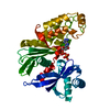
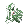
- PDB-7re6: Crystal Structure of the brown dog tick (Rhipicephalus sanguineus... -
+ Open data

- Basic information
| Entry | Database: PDB / ID: 7re6 | ||||||
|---|---|---|---|---|---|---|---|
| Title | Crystal Structure of the brown dog tick (Rhipicephalus sanguineus) Arginine Kinase in absence of substrate or ligands | ||||||
 Components Components | Arginine kinase | ||||||
 Keywords Keywords | TRANSFERASE / phosphotransferase / monomer / phosphagen | ||||||
| Biological species |  Rhipicephalus sanguineus (brown dog tick) Rhipicephalus sanguineus (brown dog tick) | ||||||
| Method |  X-RAY DIFFRACTION / SYNCHROTRON / MOLECULAR REPLACEMENT / Resolution: 1.53 Å X-RAY DIFFRACTION / SYNCHROTRON / MOLECULAR REPLACEMENT / Resolution: 1.53 Å | ||||||
 Authors Authors | Gomez-Yanes, A.C. / Garcia-Orozco, K.D. / Lopez-Zavala, A.A. / Sotelo-Mundo, R.R. | ||||||
| Funding support |  Mexico, 1items Mexico, 1items
| ||||||
 Citation Citation | Journal: Catalysts / Year: 2022 Title: The Arginine Kinase from the Tick Rhipicephalus sanguineus Is an Efficient Biocatalyst Authors: Gomez-Yanes, A.C. / Moreno-Cordova, E.N. / Garcia-Orozco, K.D. / Laino, A. / Islas-Osuna, M.A. / Lopez-Zavala, A.A. / Valenzuela, J.G. / Sotelo-Mundo, R.R. | ||||||
| History |
|
- Structure visualization
| Structure viewer | Molecule:  MolmilJmol/JSmol MolmilJmol/JSmol |
|---|
- Downloads & links
-Download
| PDBx/mmCIF format | 7re6.cif.gz | 183.6 KB | Display | PDBx/mmCIF format |
|---|---|---|---|---|
| PDB format | pdb7re6.ent.gz | 116.2 KB | Display | PDB format |
| PDBx/mmJSON format | 7re6.json.gz | Tree view | PDBx/mmJSON format | |
| Others |  Other downloads Other downloads |
-Validation report
| Arichive directory | https://data.pdbj.org/pub/pdb/validation_reports/re/7re6ftp://data.pdbj.org/pub/pdb/validation_reports/re/7re6 | HTTPS FTP |
|---|
-Related structure data
| Related structure data | 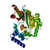 4am1S S: Starting model for refinement |
|---|---|
| Similar structure data |


-Links
 PDBj
PDBj- Assembly
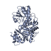
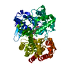
| Deposited unit | 
| ||||||||||||
|---|---|---|---|---|---|---|---|---|---|---|---|---|---|
| 1 |
| ||||||||||||
| Unit cell |
|
-Components
| #1: Protein | Mass: 42327.062 Da / Num. of mol.: 1 Source method: isolated from a genetically manipulated source Source: (gene. exp.) Rhipicephalus sanguineus (brown dog tick)Production host:  |
|---|---|
| #2: Water | ChemComp-HOH /  Mass: 18.015 Da / Num. of mol.: 377 / Source method: isolated from a natural source / Formula: H2O Mass: 18.015 Da / Num. of mol.: 377 / Source method: isolated from a natural source / Formula: H2O |
-Experimental details
-Experiment
| Experiment | Method: X-RAY DIFFRACTION / Number of used crystals: 1 |
|---|
- Sample preparation
| Crystal | Density Matthews: 2.25 Å3/Da / Density % sol: 45.23 % |
|---|---|
| Crystal grow | Temperature: 289 K / Method: microbatch / pH: 7.8 / Details: 0.1 M bis-tris pH 6.5, 25% p/v PEG 3350 |
-Data collection
| Diffraction | Mean temperature: 100 K / Serial crystal experiment: N |
|---|---|
| Diffraction source | Source: SYNCHROTRON / Site: SSRL  / Beamline: BL14-1 / Wavelength: 1.19 Å / Beamline: BL14-1 / Wavelength: 1.19 Å |
| Detector | Type: DECTRIS EIGER R 1M / Detector: PIXEL / Date: Mar 21, 2019 |
| Radiation | Protocol: SINGLE WAVELENGTH / Monochromatic (M) / Laue (L): M / Scattering type: x-ray |
| Radiation wavelength | Wavelength: 1.19 Å / Relative weight: 1 |
| Reflection | Resolution: 1.53→35.82 Å / Num. obs: 52892 / % possible obs: 98 % / Redundancy: 1.9 % / Biso Wilson estimate: 18.28 Å2 / CC1/2: 0.887 / Net I/σ(I): 8.38 |
| Reflection shell | Resolution: 1.53→1.58 Å / Num. unique obs: 52890 / CC1/2: 0.887 / % possible all: 93.01 |
- Processing
| Software |
| |||||||||||||||||||||||||||||||||||||||||||||||||||||||||||||||||||||||||||||||||||||||||||||||||||||||||||||||||||||||||||||||||||||
|---|---|---|---|---|---|---|---|---|---|---|---|---|---|---|---|---|---|---|---|---|---|---|---|---|---|---|---|---|---|---|---|---|---|---|---|---|---|---|---|---|---|---|---|---|---|---|---|---|---|---|---|---|---|---|---|---|---|---|---|---|---|---|---|---|---|---|---|---|---|---|---|---|---|---|---|---|---|---|---|---|---|---|---|---|---|---|---|---|---|---|---|---|---|---|---|---|---|---|---|---|---|---|---|---|---|---|---|---|---|---|---|---|---|---|---|---|---|---|---|---|---|---|---|---|---|---|---|---|---|---|---|---|---|---|
| Refinement | Method to determine structure: MOLECULAR REPLACEMENT Starting model: 4AM1 Resolution: 1.53→35.82 Å / SU ML: 0.1319 / Cross valid method: FREE R-VALUE / σ(F): 1.34 / Phase error: 17.9367 Stereochemistry target values: GeoStd + Monomer Library + CDL v1.2
| |||||||||||||||||||||||||||||||||||||||||||||||||||||||||||||||||||||||||||||||||||||||||||||||||||||||||||||||||||||||||||||||||||||
| Solvent computation | Shrinkage radii: 0.9 Å / VDW probe radii: 1.11 Å / Solvent model: FLAT BULK SOLVENT MODEL | |||||||||||||||||||||||||||||||||||||||||||||||||||||||||||||||||||||||||||||||||||||||||||||||||||||||||||||||||||||||||||||||||||||
| Displacement parameters | Biso mean: 23.24 Å2 | |||||||||||||||||||||||||||||||||||||||||||||||||||||||||||||||||||||||||||||||||||||||||||||||||||||||||||||||||||||||||||||||||||||
| Refinement step | Cycle: LAST / Resolution: 1.53→35.82 Å
| |||||||||||||||||||||||||||||||||||||||||||||||||||||||||||||||||||||||||||||||||||||||||||||||||||||||||||||||||||||||||||||||||||||
| Refine LS restraints |
| |||||||||||||||||||||||||||||||||||||||||||||||||||||||||||||||||||||||||||||||||||||||||||||||||||||||||||||||||||||||||||||||||||||
| LS refinement shell |
|
