 Movie
Movie Controller

[English] 日本語
 Yorodumi
- PDB-5u8e: Crystal Structure of substrate-free arginine kinase from spider P... -
+ Open data

- Basic information
| Entry | Database: PDB / ID: 5u8e | |||||||||||||||
|---|---|---|---|---|---|---|---|---|---|---|---|---|---|---|---|---|
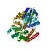
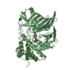
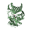
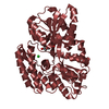
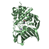
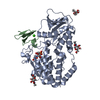
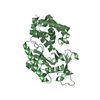
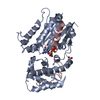
| Title | Crystal Structure of substrate-free arginine kinase from spider Polybetes pythagoricus | |||||||||||||||
 Components Components | arginine kinase | |||||||||||||||
 Keywords Keywords | TRANSFERASE / phosphagen metabolism / arginine kinase / spider / open conformation / free-ligand | |||||||||||||||
| Function / homology |  Function and homology information Function and homology informationarginine kinase / arginine kinase activity / phosphocreatine biosynthetic process / creatine kinase activity / phosphorylation / : / ATP binding Similarity search - Function | |||||||||||||||
| Biological species |  Polybetes pythagoricus (spider) Polybetes pythagoricus (spider) | |||||||||||||||
| Method |  X-RAY DIFFRACTION / MOLECULAR REPLACEMENT / Resolution: 2.18 Å X-RAY DIFFRACTION / MOLECULAR REPLACEMENT / Resolution: 2.18 Å | |||||||||||||||
 Authors Authors | Lopez-Zavala, A.A. / Garcia, C.F. / Hernadez-Paredes, J. / Sotelo-Mundo, R.R. | |||||||||||||||
| Funding support |  Mexico, Mexico,  Argentina, 4items Argentina, 4items
| |||||||||||||||
 Citation Citation | Journal: PeerJ / Year: 2017 Title: Biochemical and structural characterization of a novel arginine kinase from the spider Polybetes pythagoricus. Authors: Laino, A. / Lopez-Zavala, A.A. / Garcia-Orozco, K.D. / Carrasco-Miranda, J.S. / Santana, M. / Stojanoff, V. / Sotelo-Mundo, R.R. / Garcia, C.F. | |||||||||||||||
| History |
|
- Structure visualization
| Structure viewer | Molecule: MolmilJmol/JSmol |
|---|
- Downloads & links
-Download
| PDBx/mmCIF format | 5u8e.cif.gz | 89.6 KB | Display | PDBx/mmCIF format |
|---|---|---|---|---|
| PDB format | pdb5u8e.ent.gz | 65.1 KB | Display | PDB format |
| PDBx/mmJSON format | 5u8e.json.gz | Tree view | PDBx/mmJSON format | |
| Others |  Other downloads Other downloads |
-Validation report
| Arichive directory | https://data.pdbj.org/pub/pdb/validation_reports/u8/5u8eftp://data.pdbj.org/pub/pdb/validation_reports/u8/5u8e | HTTPS FTP |
|---|
-Related structure data
| Related structure data | 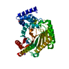 5u92C 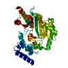 4am1S C: citing same article ( S: Starting model for refinement |
|---|---|
| Similar structure data |


-Links
 PDBj
PDBj
- Assembly
| Deposited unit | 
| ||||||||
|---|---|---|---|---|---|---|---|---|---|
| 1 |
| ||||||||
| Unit cell |
|
-Components
| #1: Protein | Mass: 43291.043 Da / Num. of mol.: 1 Source method: isolated from a genetically manipulated source Details: two loops were found disordered and Cys was oxidized form Source: (gene. exp.) Polybetes pythagoricus (spider) / Plasmid: pjexpress414 / Details (production host): T7 promoterB, Ampr, pUC originC / Production host:  |
|---|---|
| #2: Chemical | ChemComp-NA /   Mass: 22.990 Da / Num. of mol.: 1 / Source method: obtained synthetically / Formula: Na Mass: 22.990 Da / Num. of mol.: 1 / Source method: obtained synthetically / Formula: Na |
| #3: Water | ChemComp-HOH /  Mass: 18.015 Da / Num. of mol.: 266 / Source method: isolated from a natural source / Formula: H2O Mass: 18.015 Da / Num. of mol.: 266 / Source method: isolated from a natural source / Formula: H2O |
| Has protein modification | Y |
-Experimental details
-Experiment
| Experiment | Method: X-RAY DIFFRACTION / Number of used crystals: 1 |
|---|
- Sample preparation
| Crystal | Density Matthews: 2.23 Å3/Da / Density % sol: 40.33 % / Description: large-thin plates, colorless |
|---|---|
| Crystal grow | Temperature: 289 K / Method: vapor diffusion, hanging drop / pH: 5.6 Details: 0.2 M ammonium acetate, 0.1 M sodium citrate tribasic dihydrate pH 5.6 and 30% w/v polyethylene glycol 4000. Temp details: none |
-Data collection
| Diffraction | Mean temperature: 100 K |
|---|---|
| Diffraction source | Source: SEALED TUBE / Type: BRUKER IMUS MICROFOCUS / Wavelength: 1.54178 Å |
| Detector | Type: BRUKER PHOTON 100 / Detector: PIXEL / Date: Apr 27, 2016 / Details: quazar multilayer |
| Radiation | Monochromator: sealed tube/flat / Protocol: SINGLE WAVELENGTH / Monochromatic (M) / Laue (L): M / Scattering type: x-ray |
| Radiation wavelength | Wavelength: 1.54178 Å / Relative weight: 1 |
| Reflection | Resolution: 2.18→20.3 Å / Num. obs: 18342 / % possible obs: 99.43 % / Redundancy: 6.8 % / Biso Wilson estimate: 19.38 Å2 / CC1/2: 0.98 / Rmerge(I) obs: 0.0476 / Rsym value: 0.0847 / Net I/σ(I): 24.7 |
| Reflection shell | Resolution: 2.18→2.25 Å / Redundancy: 4.3 % / Rmerge(I) obs: 0.284 / Mean I/σ(I) obs: 3.34 / CC1/2: 0.739 / % possible all: 94.27 |
- Processing
| Software |
| ||||||||||||||||||||||||||||||||||||||||||||||||||||||||
|---|---|---|---|---|---|---|---|---|---|---|---|---|---|---|---|---|---|---|---|---|---|---|---|---|---|---|---|---|---|---|---|---|---|---|---|---|---|---|---|---|---|---|---|---|---|---|---|---|---|---|---|---|---|---|---|---|---|
| Refinement | Method to determine structure: MOLECULAR REPLACEMENT Starting model: 4AM1 Resolution: 2.18→20.307 Å / SU ML: 0.21 / Cross valid method: FREE R-VALUE / σ(F): 1.34 / Phase error: 22.44 / Stereochemistry target values: ML
| ||||||||||||||||||||||||||||||||||||||||||||||||||||||||
| Solvent computation | Shrinkage radii: 0.9 Å / VDW probe radii: 1.11 Å / Solvent model: FLAT BULK SOLVENT MODEL | ||||||||||||||||||||||||||||||||||||||||||||||||||||||||
| Refinement step | Cycle: LAST / Resolution: 2.18→20.307 Å
| ||||||||||||||||||||||||||||||||||||||||||||||||||||||||
| Refine LS restraints |
| ||||||||||||||||||||||||||||||||||||||||||||||||||||||||
| LS refinement shell |
|
