 Movie
Movie Controller

[English] 日本語
 Yorodumi
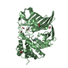
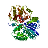
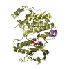
- PDB-6iff: Crystal structure of M1 zinc metallopeptidase E323A mutant from D... -
+ Open data

- Basic information
| Entry | Database: PDB / ID: 6iff | ||||||
|---|---|---|---|---|---|---|---|
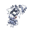
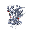
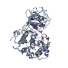
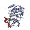
| Title | Crystal structure of M1 zinc metallopeptidase E323A mutant from Deinococcus radiodurans | ||||||
 Components Components | Zinc metalloprotease | ||||||
 Keywords Keywords | HYDROLASE / Metalloprotease / peptidase | ||||||
| Function / homology |  Function and homology information Function and homology informationmembrane alanyl aminopeptidase / alanyl aminopeptidase activity / metallopeptidase activity / proteolysis / zinc ion binding / cytoplasm Similarity search - Function | ||||||
| Biological species |  Deinococcus radiodurans (radioresistant) Deinococcus radiodurans (radioresistant) | ||||||
| Method |  X-RAY DIFFRACTION / SYNCHROTRON / MOLECULAR REPLACEMENT / Resolution: 1.83 Å X-RAY DIFFRACTION / SYNCHROTRON / MOLECULAR REPLACEMENT / Resolution: 1.83 Å | ||||||
 Authors Authors | Agrawal, R. / Kumar, A. / Kumar, A. / Gaur, N.K. / Makde, R.D. | ||||||
 Citation Citation | Journal: Int.J.Biol.Macromol. / Year: 2020 Title: Structural basis for the unusual substrate specificity of unique two-domain M1 metallopeptidase. Authors: Agrawal, R. / Goyal, V.D. / Singh, R. / Kumar, A. / Jamdar, S.N. / Kumar, A. / Makde, R.D. | ||||||
| History |
|
- Structure visualization
| Structure viewer | Molecule: MolmilJmol/JSmol |
|---|
- Downloads & links
-Download
| PDBx/mmCIF format | 6iff.cif.gz | 344.8 KB | Display | PDBx/mmCIF format |
|---|---|---|---|---|
| PDB format | pdb6iff.ent.gz | 279.1 KB | Display | PDB format |
| PDBx/mmJSON format | 6iff.json.gz | Tree view | PDBx/mmJSON format | |
| Others |  Other downloads Other downloads |
-Validation report
| Arichive directory | https://data.pdbj.org/pub/pdb/validation_reports/if/6iffftp://data.pdbj.org/pub/pdb/validation_reports/if/6iff | HTTPS FTP |
|---|
-Related structure data
| Related structure data | 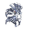 6koyC 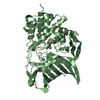 6kozC 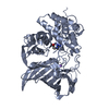 6kp0C 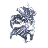 6kp1C 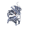 6a8zS S: Starting model for refinement C: citing same article ( |
|---|---|
| Similar structure data |


-Links
 PDBj
PDBj

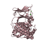
- Assembly
| Deposited unit | 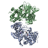
| ||||||||
|---|---|---|---|---|---|---|---|---|---|
| 1 | 
| ||||||||
| 2 | 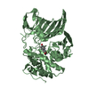
| ||||||||
| Unit cell |
|
-Components
| #1: Protein | Mass: 51547.793 Da / Num. of mol.: 2 / Mutation: E323A Source method: isolated from a genetically manipulated source Source: (gene. exp.) Deinococcus radiodurans (strain ATCC 13939 / DSM 20539 / JCM 16871 / LMG 4051 / NBRC 15346 / NCIMB 9279 / R1 / VKM B-1422) (radioresistant)Strain: ATCC 13939 / DSM 20539 / JCM 16871 / LMG 4051 / NBRC 15346 / NCIMB 9279 / R1 / VKM B-1422 Gene: DR_0875 / Plasmid: pST50TR / Details (production host): pET3a based / Production host: #2: Chemical |   Mass: 65.409 Da / Num. of mol.: 2 / Source method: obtained synthetically / Formula: Zn / Feature type: SUBJECT OF INVESTIGATION Mass: 65.409 Da / Num. of mol.: 2 / Source method: obtained synthetically / Formula: Zn / Feature type: SUBJECT OF INVESTIGATION#3: Chemical |   Type: L-peptide linking / Mass: 181.189 Da / Num. of mol.: 2 / Source method: obtained synthetically / Formula: C9H11NO3 / Feature type: SUBJECT OF INVESTIGATION Type: L-peptide linking / Mass: 181.189 Da / Num. of mol.: 2 / Source method: obtained synthetically / Formula: C9H11NO3 / Feature type: SUBJECT OF INVESTIGATION#4: Chemical |   Mass: 22.990 Da / Num. of mol.: 2 / Source method: obtained synthetically / Formula: Na Mass: 22.990 Da / Num. of mol.: 2 / Source method: obtained synthetically / Formula: Na#5: Water | ChemComp-HOH / |  Mass: 18.015 Da / Num. of mol.: 436 / Source method: isolated from a natural source / Formula: H2O Mass: 18.015 Da / Num. of mol.: 436 / Source method: isolated from a natural source / Formula: H2O |
|---|
-Experimental details
-Experiment
| Experiment | Method: X-RAY DIFFRACTION / Number of used crystals: 1 |
|---|
- Sample preparation
| Crystal | Density Matthews: 1.88 Å3/Da / Density % sol: 34.54 % / Description: Plate like crystal |
|---|---|
| Crystal grow | Temperature: 294 K / Method: microbatch / pH: 5.5 / Details: 0.2M ammonium formate, 0.1M Bis-tris, 20% PEG 3350 / PH range: 5.0-7.0 |
-Data collection
| Diffraction | Mean temperature: 100 K |
|---|---|
| Diffraction source | Source: SYNCHROTRON / Site: RRCAT INDUS-2  / Beamline: PX-BL21 / Wavelength: 0.97947 Å / Beamline: PX-BL21 / Wavelength: 0.97947 Å |
| Detector | Type: MAR scanner 345 mm plate / Detector: IMAGE PLATE / Date: Jul 27, 2018 / Details: Mirrors |
| Radiation | Monochromator: Si (111) / Protocol: SINGLE WAVELENGTH / Monochromatic (M) / Laue (L): M / Scattering type: x-ray |
| Radiation wavelength | Wavelength: 0.97947 Å / Relative weight: 1 |
| Reflection | Resolution: 1.83→45.36 Å / Num. obs: 64195 / % possible obs: 97.3 % / Redundancy: 2.5 % / Biso Wilson estimate: 8.397 Å2 / CC1/2: 0.994 / Rmerge(I) obs: 0.065 / Rpim(I) all: 0.053 / Rrim(I) all: 0.084 / Net I/σ(I): 12.2 |
| Reflection shell | Resolution: 1.83→1.87 Å / Redundancy: 2 % / Rmerge(I) obs: 0.49 / Mean I/σ(I) obs: 1.9 / Num. unique obs: 3730 / CC1/2: 0.657 / Rpim(I) all: 0.415 / Rrim(I) all: 0.645 / % possible all: 89.9 |
- Processing
| Software |
| |||||||||||||||||||||||||||||||||||||||||||||||||||||||||||||||||||||||||||||||||||||||||||||||||||||||||||||||||||||||||||||||||||||||||||||||||||||||||||||||||
|---|---|---|---|---|---|---|---|---|---|---|---|---|---|---|---|---|---|---|---|---|---|---|---|---|---|---|---|---|---|---|---|---|---|---|---|---|---|---|---|---|---|---|---|---|---|---|---|---|---|---|---|---|---|---|---|---|---|---|---|---|---|---|---|---|---|---|---|---|---|---|---|---|---|---|---|---|---|---|---|---|---|---|---|---|---|---|---|---|---|---|---|---|---|---|---|---|---|---|---|---|---|---|---|---|---|---|---|---|---|---|---|---|---|---|---|---|---|---|---|---|---|---|---|---|---|---|---|---|---|---|---|---|---|---|---|---|---|---|---|---|---|---|---|---|---|---|---|---|---|---|---|---|---|---|---|---|---|---|---|---|---|---|
| Refinement | Method to determine structure: MOLECULAR REPLACEMENT Starting model: 6A8Z Resolution: 1.83→22.265 Å / SU ML: 0.21 / Cross valid method: FREE R-VALUE / σ(F): 1.96 / Phase error: 21.67
| |||||||||||||||||||||||||||||||||||||||||||||||||||||||||||||||||||||||||||||||||||||||||||||||||||||||||||||||||||||||||||||||||||||||||||||||||||||||||||||||||
| Solvent computation | Shrinkage radii: 0.9 Å / VDW probe radii: 1.11 Å | |||||||||||||||||||||||||||||||||||||||||||||||||||||||||||||||||||||||||||||||||||||||||||||||||||||||||||||||||||||||||||||||||||||||||||||||||||||||||||||||||
| Refinement step | Cycle: LAST / Resolution: 1.83→22.265 Å
| |||||||||||||||||||||||||||||||||||||||||||||||||||||||||||||||||||||||||||||||||||||||||||||||||||||||||||||||||||||||||||||||||||||||||||||||||||||||||||||||||
| Refine LS restraints |
| |||||||||||||||||||||||||||||||||||||||||||||||||||||||||||||||||||||||||||||||||||||||||||||||||||||||||||||||||||||||||||||||||||||||||||||||||||||||||||||||||
| LS refinement shell |
| |||||||||||||||||||||||||||||||||||||||||||||||||||||||||||||||||||||||||||||||||||||||||||||||||||||||||||||||||||||||||||||||||||||||||||||||||||||||||||||||||
| Refinement TLS params. | Method: refined / Origin x: -41.5789 Å / Origin y: -41.0229 Å / Origin z: 6.007 Å
| |||||||||||||||||||||||||||||||||||||||||||||||||||||||||||||||||||||||||||||||||||||||||||||||||||||||||||||||||||||||||||||||||||||||||||||||||||||||||||||||||
| Refinement TLS group | Selection details: all |
