 Movie
Movie Controller

[English] 日本語
 Yorodumi
- PDB-6koz: Crystal structure of two domain M1 zinc metallopeptidase E323 mut... -
+ Open data

- Basic information
| Entry | Database: PDB / ID: 6koz | ||||||
|---|---|---|---|---|---|---|---|
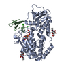
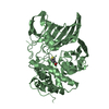
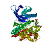
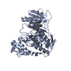
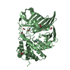
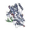
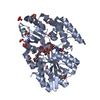
| Title | Crystal structure of two domain M1 zinc metallopeptidase E323 mutant bound to L-Leucine amino acid | ||||||
 Components Components | Zinc metalloprotease, putative | ||||||
 Keywords Keywords | HYDROLASE / Metalloprotease | ||||||
| Function / homology |  Function and homology information Function and homology informationmembrane alanyl aminopeptidase / alanyl aminopeptidase activity / metallopeptidase activity / proteolysis / zinc ion binding / cytoplasm Similarity search - Function | ||||||
| Biological species |  Deinococcus radiodurans (radioresistant) Deinococcus radiodurans (radioresistant) | ||||||
| Method |  X-RAY DIFFRACTION / SYNCHROTRON / MOLECULAR REPLACEMENT / Resolution: 2.25 Å X-RAY DIFFRACTION / SYNCHROTRON / MOLECULAR REPLACEMENT / Resolution: 2.25 Å | ||||||
 Authors Authors | Agrawal, R. / Kumar, A. / Kumar, A. / Makde, R.D. | ||||||
 Citation Citation | Journal: Int.J.Biol.Macromol. / Year: 2020 Title: Structural basis for the unusual substrate specificity of unique two-domain M1 metallopeptidase. Authors: Agrawal, R. / Goyal, V.D. / Singh, R. / Kumar, A. / Jamdar, S.N. / Kumar, A. / Makde, R.D. | ||||||
| History |
|
- Structure visualization
| Structure viewer | Molecule: MolmilJmol/JSmol |
|---|
- Downloads & links
-Download
| PDBx/mmCIF format | 6koz.cif.gz | 186.7 KB | Display | PDBx/mmCIF format |
|---|---|---|---|---|
| PDB format | pdb6koz.ent.gz | 144.1 KB | Display | PDB format |
| PDBx/mmJSON format | 6koz.json.gz | Tree view | PDBx/mmJSON format | |
| Others |  Other downloads Other downloads |
-Validation report
| Arichive directory | https://data.pdbj.org/pub/pdb/validation_reports/ko/6kozftp://data.pdbj.org/pub/pdb/validation_reports/ko/6koz | HTTPS FTP |
|---|
-Related structure data
| Related structure data |  6iffC 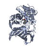 6koyC 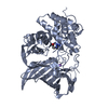 6kp0C 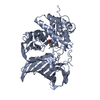 6kp1C 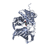 6a8zS S: Starting model for refinement C: citing same article ( |
|---|---|
| Similar structure data |


-Links
 PDBj
PDBj


- Assembly
| Deposited unit | 
| ||||||||||||||||||||||||||||||||||||||||||||||||||||||||||||||||||||||||||||||||||||||||||
|---|---|---|---|---|---|---|---|---|---|---|---|---|---|---|---|---|---|---|---|---|---|---|---|---|---|---|---|---|---|---|---|---|---|---|---|---|---|---|---|---|---|---|---|---|---|---|---|---|---|---|---|---|---|---|---|---|---|---|---|---|---|---|---|---|---|---|---|---|---|---|---|---|---|---|---|---|---|---|---|---|---|---|---|---|---|---|---|---|---|---|---|
| 1 | 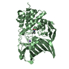
| ||||||||||||||||||||||||||||||||||||||||||||||||||||||||||||||||||||||||||||||||||||||||||
| 2 | 
| ||||||||||||||||||||||||||||||||||||||||||||||||||||||||||||||||||||||||||||||||||||||||||
| Unit cell |
| ||||||||||||||||||||||||||||||||||||||||||||||||||||||||||||||||||||||||||||||||||||||||||
| Noncrystallographic symmetry (NCS) | NCS domain:
NCS domain segments: Ens-ID: 1
|