 Movie
Movie Controller

[English] 日本語
 Yorodumi
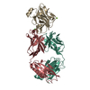
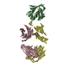
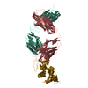
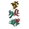
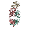
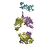
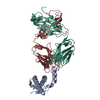
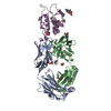
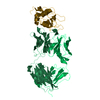
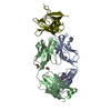
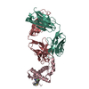
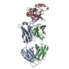
- PDB-7qu2: Junin virus GP1 glycoprotein in complex with Fab fragment of anti... -
+ Open data

- Basic information
| Entry | Database: PDB / ID: 7qu2 | |||||||||||||||||||||||||||
|---|---|---|---|---|---|---|---|---|---|---|---|---|---|---|---|---|---|---|---|---|---|---|---|---|---|---|---|---|
| Title | Junin virus GP1 glycoprotein in complex with Fab fragment of antibody JUN1 | |||||||||||||||||||||||||||
 Components Components |
| |||||||||||||||||||||||||||
 Keywords Keywords | IMMUNE SYSTEM / Arenavirus / Glycoprotein / Monoclonal Antibody / Complex | |||||||||||||||||||||||||||
| Function / homology |  Function and homology information Function and homology informationhost cell Golgi membrane / receptor-mediated endocytosis of virus by host cell / host cell endoplasmic reticulum membrane / fusion of virus membrane with host endosome membrane / viral envelope / virion attachment to host cell / host cell plasma membrane / virion membrane / metal ion binding / membrane Similarity search - Function | |||||||||||||||||||||||||||
| Biological species |   Argentinian mammarenavirus Argentinian mammarenavirus | |||||||||||||||||||||||||||
| Method |  X-RAY DIFFRACTION / SYNCHROTRON / MOLECULAR REPLACEMENT / Resolution: 2.5 Å X-RAY DIFFRACTION / SYNCHROTRON / MOLECULAR REPLACEMENT / Resolution: 2.5 Å | |||||||||||||||||||||||||||
 Authors Authors | Ng, W.M. / Sahin, M. / Krumm, S.A. / Seow, J. / Zeltina, A. / Harlos, K. / Paesen, G. / Pinschewer, D.D. / Doores, K.J. / Bowden, T.A. | |||||||||||||||||||||||||||
| Funding support |  United Kingdom, 8items United Kingdom, 8items
| |||||||||||||||||||||||||||
 Citation Citation | Journal: Mbio / Year: 2022 Title: Contrasting Modes of New World Arenavirus Neutralization by Immunization-Elicited Monoclonal Antibodies. Authors: Ng, W.M. / Sahin, M. / Krumm, S.A. / Seow, J. / Zeltina, A. / Harlos, K. / Paesen, G.C. / Pinschewer, D.D. / Doores, K.J. / Bowden, T.A. | |||||||||||||||||||||||||||
| History |
|
- Structure visualization
| Structure viewer | Molecule: MolmilJmol/JSmol |
|---|
- Downloads & links
-Download
| PDBx/mmCIF format | 7qu2.cif.gz | 252.8 KB | Display | PDBx/mmCIF format |
|---|---|---|---|---|
| PDB format | pdb7qu2.ent.gz | 200.7 KB | Display | PDB format |
| PDBx/mmJSON format | 7qu2.json.gz | Tree view | PDBx/mmJSON format | |
| Others |  Other downloads Other downloads |
-Validation report
| Arichive directory | https://data.pdbj.org/pub/pdb/validation_reports/qu/7qu2ftp://data.pdbj.org/pub/pdb/validation_reports/qu/7qu2 | HTTPS FTP |
|---|
-Related structure data
| Related structure data | 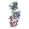 7qu1C  5nuzS S: Starting model for refinement C: citing same article ( |
|---|---|
| Similar structure data |

-Links
 PDBj
PDBj

- Assembly
| Deposited unit | 
| ||||||||
|---|---|---|---|---|---|---|---|---|---|
| 1 |
| ||||||||
| Unit cell |
|
-Components
-Protein , 1 types, 1 molecules C
| #3: Protein | Mass: 18095.586 Da / Num. of mol.: 1 Source method: isolated from a genetically manipulated source Source: (gene. exp.) Argentinian mammarenavirus / Gene: GPC / Production host:  Homo sapiens (human) / References: UniProt: C1K9J9 Homo sapiens (human) / References: UniProt: C1K9J9 |
|---|
-Antibody , 2 types, 2 molecules AB
| #1: Antibody | Mass: 26402.504 Da / Num. of mol.: 1 Source method: isolated from a genetically manipulated source Source: (gene. exp.) Homo sapiens (human) |
|---|---|
| #2: Antibody | Mass: 23943.455 Da / Num. of mol.: 1 Source method: isolated from a genetically manipulated source Source: (gene. exp.) Homo sapiens (human) |
-Sugars , 2 types, 2 molecules 
| #4: Polysaccharide | 2-acetamido-2-deoxy-beta-D-glucopyranose-(1-4)-2-acetamido-2-deoxy-beta-D-glucopyranose Source method: isolated from a genetically manipulated source |
|---|---|
| #6: Sugar | ChemComp-NAG /  Type: D-saccharide, beta linking / Mass: 221.208 Da / Num. of mol.: 1 / Source method: obtained synthetically / Formula: C8H15NO6 Type: D-saccharide, beta linking / Mass: 221.208 Da / Num. of mol.: 1 / Source method: obtained synthetically / Formula: C8H15NO6 |
-Non-polymers , 2 types, 39 molecules 

| #5: Chemical |  Mass: 92.094 Da / Num. of mol.: 2 / Source method: obtained synthetically / Formula: C3H8O3 Mass: 92.094 Da / Num. of mol.: 2 / Source method: obtained synthetically / Formula: C3H8O3#7: Water | ChemComp-HOH / | Mass: 18.015 Da / Num. of mol.: 37 / Source method: isolated from a natural source / Formula: H2O |
|---|
-Details
| Has ligand of interest | N |
|---|---|
| Has protein modification | Y |
-Experimental details
-Experiment
| Experiment | Method: X-RAY DIFFRACTION / Number of used crystals: 1 |
|---|
- Sample preparation
| Crystal | Density Matthews: 2.37 Å3/Da / Density % sol: 48.07 % |
|---|---|
| Crystal grow | Temperature: 293 K / Method: vapor diffusion, sitting drop / pH: 7.5 / Details: 20% w/v PEG8000 and 0.1 M HEPES (pH 7.5) |
-Data collection
| Diffraction | Mean temperature: 100 K / Serial crystal experiment: N |
|---|---|
| Diffraction source | Source: SYNCHROTRON / Site: Diamond / Beamline: I03 / Wavelength: 0.9686 Å |
| Detector | Type: DECTRIS PILATUS 6M / Detector: PIXEL / Date: Mar 13, 2018 |
| Radiation | Protocol: SINGLE WAVELENGTH / Monochromatic (M) / Laue (L): M / Scattering type: x-ray |
| Radiation wavelength | Wavelength: 0.9686 Å / Relative weight: 1 |
| Reflection | Resolution: 2.5→67.92 Å / Num. obs: 22859 / % possible obs: 99.8 % / Redundancy: 6.5 % / Biso Wilson estimate: 58.385 Å2 / CC1/2: 0.995 / Rmerge(I) obs: 0.131 / Net I/σ(I): 7.1 |
| Reflection shell | Resolution: 2.5→2.54 Å / Redundancy: 5.9 % / Rmerge(I) obs: 0.794 / Num. unique obs: 1100 / CC1/2: 0.742 / % possible all: 96.2 |
- Processing
| Software |
| |||||||||||||||||||||||||||||||||||||||||||||||||||||||||||||||
|---|---|---|---|---|---|---|---|---|---|---|---|---|---|---|---|---|---|---|---|---|---|---|---|---|---|---|---|---|---|---|---|---|---|---|---|---|---|---|---|---|---|---|---|---|---|---|---|---|---|---|---|---|---|---|---|---|---|---|---|---|---|---|---|---|
| Refinement | Method to determine structure: MOLECULAR REPLACEMENT Starting model: 5NUZ Resolution: 2.5→67.92 Å / SU ML: 0.38 / Cross valid method: THROUGHOUT / σ(F): 1.37 / Phase error: 33.7 / Stereochemistry target values: ML
| |||||||||||||||||||||||||||||||||||||||||||||||||||||||||||||||
| Solvent computation | Shrinkage radii: 0.9 Å / VDW probe radii: 1.11 Å / Solvent model: FLAT BULK SOLVENT MODEL | |||||||||||||||||||||||||||||||||||||||||||||||||||||||||||||||
| Displacement parameters | Biso max: 157.47 Å2 / Biso mean: 75.0604 Å2 / Biso min: 40.76 Å2 | |||||||||||||||||||||||||||||||||||||||||||||||||||||||||||||||
| Refinement step | Cycle: final / Resolution: 2.5→67.92 Å
| |||||||||||||||||||||||||||||||||||||||||||||||||||||||||||||||
| LS refinement shell | Refine-ID: X-RAY DIFFRACTION / Rfactor Rfree error: 0 / Total num. of bins used: 8
| |||||||||||||||||||||||||||||||||||||||||||||||||||||||||||||||
| Refinement TLS params. | Method: refined / Origin x: 14.1813 Å / Origin y: -0.8831 Å / Origin z: 22.3838 Å
| |||||||||||||||||||||||||||||||||||||||||||||||||||||||||||||||
| Refinement TLS group |
|
