 Movie
Movie Controller

[English] 日本語
 Yorodumi
- PDB-1bql: STRUCTURE OF AN ANTI-HEL FAB FRAGMENT COMPLEXED WITH BOBWHITE QUA... -
+ Open data

- Basic information
| Entry | Database: PDB / ID: 1bql | |||||||||
|---|---|---|---|---|---|---|---|---|---|---|
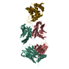
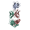
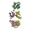
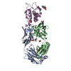
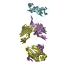
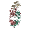
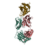
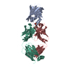
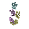
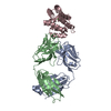
| Title | STRUCTURE OF AN ANTI-HEL FAB FRAGMENT COMPLEXED WITH BOBWHITE QUAIL LYSOZYME | |||||||||
 Components Components |
| |||||||||
 Keywords Keywords | ANTIBODY/ANTIGEN / ANTIBODY-ANTIGEN complex / HYHEL-5 / ANTI-LYSOZYME / BOBWHITE QUAIL | |||||||||
| Function / homology |  Function and homology information Function and homology informationcell wall macromolecule catabolic process / lysozyme / lysozyme activity / killing of cells of another organism / defense response to Gram-negative bacterium / defense response to Gram-positive bacterium / extracellular region / cytoplasm Similarity search - Function | |||||||||
| Biological species |   Colinus virginianus (northern bobwhite) Colinus virginianus (northern bobwhite) | |||||||||
| Method |  X-RAY DIFFRACTION / Resolution: 2.6 Å X-RAY DIFFRACTION / Resolution: 2.6 Å | |||||||||
 Authors Authors | Chacko, S. / Davies, D.R. | |||||||||
 Citation Citation | Journal: Proteins / Year: 1996 Title: Refined structures of bobwhite quail lysozyme uncomplexed and complexed with the HyHEL-5 Fab fragment. Authors: Chacko, S. / Silverton, E.W. / Smith-Gill, S.J. / Davies, D.R. / Shick, K.A. / Xavier, K.A. / Willson, R.C. / Jeffrey, P.D. / Chang, C.Y. / Sieker, L.C. / Sheriff, S. #1: Journal: J. Mol. Biol. / Year: 1995Title: Structure of an antibody-lysozyme complex unexpected effect of conservative mutation. Authors: Chacko, S. / Silverton, E. / Kam-Morgan, L. / Smith-Gill, S. / Cohen, G. / Davies, D. #2: Journal: Proc. Natl. Acad. Sci. U.S.A. / Year: 1987 Title: Three-dimensional structure of an antibody-antigen complex. Authors: Sheriff, S. / Silverton, E.W. / Padlan, E.A. / Cohen, G.H. / Smith-Gill, S.J. / Finzel, B.C. / Davies, D.R. | |||||||||
| History |
|
- Structure visualization
| Structure viewer | Molecule: MolmilJmol/JSmol |
|---|
- Downloads & links
-Download
| PDBx/mmCIF format | 1bql.cif.gz | 121.3 KB | Display | PDBx/mmCIF format |
|---|---|---|---|---|
| PDB format | pdb1bql.ent.gz | 92.8 KB | Display | PDB format |
| PDBx/mmJSON format | 1bql.json.gz | Tree view | PDBx/mmJSON format | |
| Others |  Other downloads Other downloads |
-Validation report
| Arichive directory | https://data.pdbj.org/pub/pdb/validation_reports/bq/1bqlftp://data.pdbj.org/pub/pdb/validation_reports/bq/1bql | HTTPS FTP |
|---|
-Related structure data


-Links
 PDBj
PDBj

- Assembly
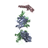
| Deposited unit | 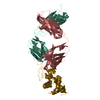
| ||||||||
|---|---|---|---|---|---|---|---|---|---|
| 1 |
| ||||||||
| Unit cell |
| ||||||||
| Atom site foot note | 1: CIS PROLINE - PRO L 8 / 2: CIS PROLINE - PRO L 139 / 3: CIS PROLINE - PRO H 150 / 4: CIS PROLINE - PRO H 152 / 5: CIS PROLINE - PRO H 192 |
-Components
| #1: Antibody | Mass: 23326.729 Da / Num. of mol.: 1 Source method: isolated from a genetically manipulated source Source: (gene. exp.) |
|---|---|
| #2: Antibody | Mass: 22948.527 Da / Num. of mol.: 1 Source method: isolated from a genetically manipulated source Source: (gene. exp.) |
| #3: Protein | Mass: 14289.119 Da / Num. of mol.: 1 Source method: isolated from a genetically manipulated source Source: (gene. exp.) Colinus virginianus (northern bobwhite)References: UniProt: P00700 |
| #4: Water | ChemComp-HOH /  Mass: 18.015 Da / Num. of mol.: 86 / Source method: isolated from a natural source / Formula: H2O Mass: 18.015 Da / Num. of mol.: 86 / Source method: isolated from a natural source / Formula: H2O |
| Compound details | COMPND FAB FRAGMENT OF AN ANTI-LYSOZYME IGG1 ANTIBODY HYHEL-5 - BOBWHITE QUAIL LYSOZYME COMPLEX. |
| Has protein modification | Y |
| Sequence details | THE LIGHT CHAIN OF THE FAB HAS THE CHAIN INDICATOR L. THE HEAVY CHAIN OF THE FAB HAS THE CHAIN ...THE LIGHT CHAIN OF THE FAB HAS THE CHAIN INDICATOR L. THE HEAVY CHAIN OF THE FAB HAS THE CHAIN INDICATOR H. THE LYSOZYME HAS THE CHAIN INDICATOR Y. THE NUMBERING SYSTEM USED IS SEQUENTIAL |
-Experimental details
-Experiment
| Experiment | Method: X-RAY DIFFRACTION |
|---|
- Sample preparation
| Crystal | Density Matthews: 2.62 Å3/Da / Density % sol: 52.98 % | ||||||||||||||||||||||||||||||||||||
|---|---|---|---|---|---|---|---|---|---|---|---|---|---|---|---|---|---|---|---|---|---|---|---|---|---|---|---|---|---|---|---|---|---|---|---|---|---|
| Crystal grow | *PLUS Method: vapor diffusion | ||||||||||||||||||||||||||||||||||||
| Components of the solutions | *PLUS
|
-Data collection
| Reflection | Num. obs: 18884 / % possible obs: 94.5 % / Observed criterion σ(I): 0 |
|---|---|
| Reflection | *PLUS Highest resolution: 2.6 Å / Num. obs: 15950 / % possible obs: 80.8 % / Num. measured all: 30801 / Rmerge(I) obs: 0.074 |
- Processing
| Software |
| ||||||||||||||||||||||||||||||||||||||||||||||||||||||||||||
|---|---|---|---|---|---|---|---|---|---|---|---|---|---|---|---|---|---|---|---|---|---|---|---|---|---|---|---|---|---|---|---|---|---|---|---|---|---|---|---|---|---|---|---|---|---|---|---|---|---|---|---|---|---|---|---|---|---|---|---|---|---|
| Refinement | Resolution: 2.6→8 Å / σ(F): 2
| ||||||||||||||||||||||||||||||||||||||||||||||||||||||||||||
| Displacement parameters | Biso mean: 22.35 Å2 | ||||||||||||||||||||||||||||||||||||||||||||||||||||||||||||
| Refine analyze | Luzzati coordinate error obs: 0.3 Å | ||||||||||||||||||||||||||||||||||||||||||||||||||||||||||||
| Refinement step | Cycle: LAST / Resolution: 2.6→8 Å
| ||||||||||||||||||||||||||||||||||||||||||||||||||||||||||||
| Refine LS restraints |
| ||||||||||||||||||||||||||||||||||||||||||||||||||||||||||||
| Refinement | *PLUS Lowest resolution: 8 Å / Num. reflection obs: 14290 / σ(F): 2 | ||||||||||||||||||||||||||||||||||||||||||||||||||||||||||||
| Solvent computation | *PLUS | ||||||||||||||||||||||||||||||||||||||||||||||||||||||||||||
| Displacement parameters | *PLUS | ||||||||||||||||||||||||||||||||||||||||||||||||||||||||||||
| Refine LS restraints | *PLUS
|
