 Movie
Movie Controller


+ Open data

- Basic information
| Entry | Database: PDB / ID: 7qs5 | ||||||
|---|---|---|---|---|---|---|---|
| Title | Crystal structure of B30.2 PRYSPRY domain of TRIM67 | ||||||
 Components Components | Tripartite motif-containing protein 67 | ||||||
 Keywords Keywords | LIGASE / E3 / TRIM / TRIM67 / B30.2 domain / SPRY domain / PRYSPRY domain | ||||||
| Function / homology |  Function and homology information Function and homology informationnegative regulation of Ras protein signal transduction / positive regulation of ubiquitin-dependent protein catabolic process / positive regulation of neuron projection development / regulation of protein localization / cytoskeleton / zinc ion binding / cytoplasm Similarity search - Function | ||||||
| Biological species |  Homo sapiens (human) Homo sapiens (human) | ||||||
| Method |  X-RAY DIFFRACTION / SYNCHROTRON / MOLECULAR REPLACEMENT / Resolution: 1.65 Å X-RAY DIFFRACTION / SYNCHROTRON / MOLECULAR REPLACEMENT / Resolution: 1.65 Å | ||||||
 Authors Authors | Chaikuad, A. / Zhubi, R. / Knapp, S. / Structural Genomics Consortium (SGC) | ||||||
| Funding support | 1items
| ||||||
 Citation Citation | Journal: J Struct Biol X / Year: 2025 Title: Structural analysis of TRIM family PRYSPRY domains and its implications for E3-ligand design. Authors: Zhubi, R. / Chaikuad, A. / Munoz Sosa, C.J. / Joerger, A.C. / Knapp, S. | ||||||
| History |
|
- Structure visualization
| Structure viewer | Molecule: MolmilJmol/JSmol |
|---|
- Downloads & links
-Download
| PDBx/mmCIF format | 7qs5.cif.gz | 149.5 KB | Display | PDBx/mmCIF format |
|---|---|---|---|---|
| PDB format | pdb7qs5.ent.gz | 117 KB | Display | PDB format |
| PDBx/mmJSON format | 7qs5.json.gz | Tree view | PDBx/mmJSON format | |
| Others |  Other downloads Other downloads |
-Validation report
| Arichive directory | https://data.pdbj.org/pub/pdb/validation_reports/qs/7qs5ftp://data.pdbj.org/pub/pdb/validation_reports/qs/7qs5 | HTTPS FTP |
|---|
-Related structure data
| Related structure data |  7b2sSC 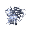 7qryC  7qrzC 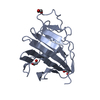 7qs0C 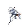 7qs1C 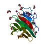 7qs2C 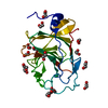 7qs3C 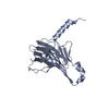 7qs4C  9r11C S: Starting model for refinement C: citing same article ( |
|---|---|
| Similar structure data |
-Links
 PDBj
PDBj


- Assembly
| Deposited unit | 
| ||||||||||||||||||
|---|---|---|---|---|---|---|---|---|---|---|---|---|---|---|---|---|---|---|---|
| 1 | 
| ||||||||||||||||||
| 2 | 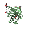
| ||||||||||||||||||
| Unit cell |
| ||||||||||||||||||
| Noncrystallographic symmetry (NCS) | NCS domain:
NCS domain segments: Component-ID: _ / Ens-ID: 1 / Beg auth comp-ID: TRP / Beg label comp-ID: TRP / End auth comp-ID: HIS / End label comp-ID: HIS / Refine code: _ / Auth seq-ID: 609 - 766 / Label seq-ID: 6 - 163
|
-Components
| #1: Protein | Mass: 19950.309 Da / Num. of mol.: 2 Source method: isolated from a genetically manipulated source Source: (gene. exp.) Homo sapiens (human) / Gene: TRIM67, TNL / Plasmid: pGTVL2 / Production host:  #2: Chemical | ChemComp-EDO /   Mass: 62.068 Da / Num. of mol.: 19 / Source method: obtained synthetically / Formula: C2H6O2 Mass: 62.068 Da / Num. of mol.: 19 / Source method: obtained synthetically / Formula: C2H6O2#3: Water | ChemComp-HOH / |  Mass: 18.015 Da / Num. of mol.: 221 / Source method: isolated from a natural source / Formula: H2O Mass: 18.015 Da / Num. of mol.: 221 / Source method: isolated from a natural source / Formula: H2OHas ligand of interest | N | Has protein modification | Y | |
|---|
-Experimental details
-Experiment
| Experiment | Method: X-RAY DIFFRACTION / Number of used crystals: 1 |
|---|
- Sample preparation
| Crystal | Density Matthews: 2.06 Å3/Da / Density % sol: 40.17 % |
|---|---|
| Crystal grow | Temperature: 293.15 K / Method: vapor diffusion, sitting drop / pH: 4.5 Details: 20% high molecular weight PEG Smears, 0.1M acetate pH 4.5 |
-Data collection
| Diffraction | Mean temperature: 100 K / Serial crystal experiment: N | |||||||||||||||||||||||||||
|---|---|---|---|---|---|---|---|---|---|---|---|---|---|---|---|---|---|---|---|---|---|---|---|---|---|---|---|---|
| Diffraction source | Source: SYNCHROTRON / Site: SLS  / Beamline: X06SA / Wavelength: 0.99981 Å / Beamline: X06SA / Wavelength: 0.99981 Å | |||||||||||||||||||||||||||
| Detector | Type: DECTRIS EIGER2 X 16M / Detector: PIXEL / Date: Oct 20, 2021 | |||||||||||||||||||||||||||
| Radiation | Protocol: SINGLE WAVELENGTH / Monochromatic (M) / Laue (L): M / Scattering type: x-ray | |||||||||||||||||||||||||||
| Radiation wavelength | Wavelength: 0.99981 Å / Relative weight: 1 | |||||||||||||||||||||||||||
| Reflection | Resolution: 1.65→47.87 Å / Num. obs: 39503 / % possible obs: 99.9 % / Redundancy: 10.6 % / CC1/2: 0.999 / Rmerge(I) obs: 0.063 / Rpim(I) all: 0.021 / Rrim(I) all: 0.069 / Net I/σ(I): 17.7 | |||||||||||||||||||||||||||
| Reflection shell | Diffraction-ID: 1
|
- Processing
| Software |
| |||||||||||||||||||||||||||||||||||||||||||||||||||||||||||||||||||||||||||
|---|---|---|---|---|---|---|---|---|---|---|---|---|---|---|---|---|---|---|---|---|---|---|---|---|---|---|---|---|---|---|---|---|---|---|---|---|---|---|---|---|---|---|---|---|---|---|---|---|---|---|---|---|---|---|---|---|---|---|---|---|---|---|---|---|---|---|---|---|---|---|---|---|---|---|---|---|
| Refinement | Method to determine structure: MOLECULAR REPLACEMENT Starting model: 7B2S Resolution: 1.65→47.87 Å / Cor.coef. Fo:Fc: 0.973 / Cor.coef. Fo:Fc free: 0.967 / SU B: 3.463 / SU ML: 0.058 / SU R Cruickshank DPI: 0.0867 / Cross valid method: THROUGHOUT / σ(F): 0 / ESU R: 0.087 / ESU R Free: 0.082 / Stereochemistry target values: MAXIMUM LIKELIHOOD Details: HYDROGENS HAVE BEEN ADDED IN THE RIDING POSITIONS U VALUES : WITH TLS ADDED
| |||||||||||||||||||||||||||||||||||||||||||||||||||||||||||||||||||||||||||
| Solvent computation | Ion probe radii: 0.8 Å / Shrinkage radii: 0.8 Å / VDW probe radii: 1.2 Å / Solvent model: MASK | |||||||||||||||||||||||||||||||||||||||||||||||||||||||||||||||||||||||||||
| Displacement parameters | Biso max: 93.17 Å2 / Biso mean: 28.266 Å2 / Biso min: 15.81 Å2
| |||||||||||||||||||||||||||||||||||||||||||||||||||||||||||||||||||||||||||
| Refinement step | Cycle: final / Resolution: 1.65→47.87 Å
| |||||||||||||||||||||||||||||||||||||||||||||||||||||||||||||||||||||||||||
| Refine LS restraints |
| |||||||||||||||||||||||||||||||||||||||||||||||||||||||||||||||||||||||||||
| Refine LS restraints NCS | Ens-ID: 1 / Number: 5257 / Refine-ID: X-RAY DIFFRACTION / Type: interatomic distance / Rms dev position: 0.1 Å / Weight position: 0.05
| |||||||||||||||||||||||||||||||||||||||||||||||||||||||||||||||||||||||||||
| LS refinement shell | Resolution: 1.65→1.693 Å / Rfactor Rfree error: 0 / Total num. of bins used: 20
| |||||||||||||||||||||||||||||||||||||||||||||||||||||||||||||||||||||||||||
| Refinement TLS params. | Method: refined / Refine-ID: X-RAY DIFFRACTION
| |||||||||||||||||||||||||||||||||||||||||||||||||||||||||||||||||||||||||||
| Refinement TLS group |
|
