 Movie
Movie Controller

[English] 日本語
 Yorodumi
- PDB-7qos: Cyclopropane fatty acid synthase from Aquifex aeolicous with boun... -
+ Open data

- Basic information
| Entry | Database: PDB / ID: 7qos | ||||||
|---|---|---|---|---|---|---|---|
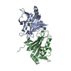
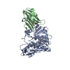
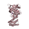
| Title | Cyclopropane fatty acid synthase from Aquifex aeolicous with bound ligands | ||||||
 Components Components | Cyclopropane-fatty-acyl-phospholipid synthase | ||||||
 Keywords Keywords | TRANSFERASE / phospholipid / complex / cyclopropane fatty acid synthase / methyl transferase / SAM | ||||||
| Function / homology |  Function and homology information Function and homology information | ||||||
| Biological species |   Aquifex aeolicus VF5 (bacteria) Aquifex aeolicus VF5 (bacteria) | ||||||
| Method |  X-RAY DIFFRACTION / SYNCHROTRON / MOLECULAR REPLACEMENT / molecular replacement / Resolution: 1.6 Å X-RAY DIFFRACTION / SYNCHROTRON / MOLECULAR REPLACEMENT / molecular replacement / Resolution: 1.6 Å | ||||||
 Authors Authors | Lukk, T. / Cronan, J.E. / Nair, S.K. | ||||||
| Funding support |  United States, 1items United States, 1items
| ||||||
 Citation Citation | Journal: To Be Published Title: Structure of cyclopropane fatty acid synthase from Aquifex aeolicus Authors: Cronan, J.E. / Nair, S.K. / Lukk, T. #1: Journal: Microbiol. Mol. Biol. Rev. / Year: 2022 Title: Advances in the Structural Biology, Mechanism, and Physiology of Cyclopropane Fatty Acid Modifications of Bacterial Membranes Authors: Cronan, J.E. / Lukk, T. | ||||||
| History |
|
- Structure visualization
| Structure viewer | Molecule: MolmilJmol/JSmol |
|---|
- Downloads & links
-Download
| PDBx/mmCIF format | 7qos.cif.gz | 178.6 KB | Display | PDBx/mmCIF format |
|---|---|---|---|---|
| PDB format | pdb7qos.ent.gz | 136.6 KB | Display | PDB format |
| PDBx/mmJSON format | 7qos.json.gz | Tree view | PDBx/mmJSON format | |
| Others |  Other downloads Other downloads |
-Validation report
| Summary document | 7qos_validation.pdf.gz | 2 MB | Display | wwPDB validaton report |
|---|---|---|---|---|
| Full document | 7qos_full_validation.pdf.gz | 2 MB | Display | |
| Data in XML | 7qos_validation.xml.gz | 33.9 KB | Display | |
| Data in CIF | 7qos_validation.cif.gz | 49.6 KB | Display | |
| Arichive directory | https://data.pdbj.org/pub/pdb/validation_reports/qo/7qosftp://data.pdbj.org/pub/pdb/validation_reports/qo/7qos | HTTPS FTP |
-Related structure data
| Related structure data | 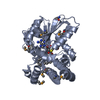 1kpgS S: Starting model for refinement |
|---|---|
| Similar structure data |


-Links
 PDBj
PDBj


- Assembly
| Deposited unit | 
| ||||||||
|---|---|---|---|---|---|---|---|---|---|
| 1 | 
| ||||||||
| 2 | 
| ||||||||
| Unit cell |
|
-Components
-Protein , 1 types, 2 molecules AB
| #1: Protein | Mass: 47487.590 Da / Num. of mol.: 2 Source method: isolated from a genetically manipulated source Source: (gene. exp.) Aquifex aeolicus VF5 (bacteria) / Strain: VF5 / Gene: cfa, aq_1737 / Plasmid: pET28 / Production host: |
|---|
-Non-polymers , 5 types, 581 molecules 


| #2: Chemical |  Mass: 675.960 Da / Num. of mol.: 2 / Source method: obtained synthetically / Formula: C37H74NO7P / Feature type: SUBJECT OF INVESTIGATION Mass: 675.960 Da / Num. of mol.: 2 / Source method: obtained synthetically / Formula: C37H74NO7P / Feature type: SUBJECT OF INVESTIGATION#3: Chemical |  Mass: 60.009 Da / Num. of mol.: 2 / Source method: obtained synthetically / Formula: CO3 / Feature type: SUBJECT OF INVESTIGATION Mass: 60.009 Da / Num. of mol.: 2 / Source method: obtained synthetically / Formula: CO3 / Feature type: SUBJECT OF INVESTIGATION#4: Chemical | ChemComp-SAH / |  Type: L-peptide linking / Mass: 384.411 Da / Num. of mol.: 1 / Source method: obtained synthetically / Formula: C14H20N6O5S / Feature type: SUBJECT OF INVESTIGATION Type: L-peptide linking / Mass: 384.411 Da / Num. of mol.: 1 / Source method: obtained synthetically / Formula: C14H20N6O5S / Feature type: SUBJECT OF INVESTIGATION#5: Chemical | ChemComp-SAM / |  Mass: 398.437 Da / Num. of mol.: 1 / Source method: obtained synthetically / Formula: C15H22N6O5S / Feature type: SUBJECT OF INVESTIGATION Mass: 398.437 Da / Num. of mol.: 1 / Source method: obtained synthetically / Formula: C15H22N6O5S / Feature type: SUBJECT OF INVESTIGATION#6: Water | ChemComp-HOH / | Mass: 18.015 Da / Num. of mol.: 575 / Source method: isolated from a natural source / Formula: H2O |
|---|
-Details
| Has ligand of interest | Y |
|---|
-Experimental details
-Experiment
| Experiment | Method: X-RAY DIFFRACTION / Number of used crystals: 1 |
|---|
- Sample preparation
| Crystal | Density Matthews: 2.36 Å3/Da / Density % sol: 47.97 % |
|---|---|
| Crystal grow | Temperature: 282 K / Method: vapor diffusion, hanging drop / pH: 7.5 Details: 0.2M L-pro, 10% PEG 3350, 0.1M HEPES 7.5, 5 mM S-adenosyl-L-Methionine; protein concentration 8 mg/mL, 1:1 ratio of protein to mother liquor. Protein storage buffer: 20 mM Tris pH 8.5, 0.5M ...Details: 0.2M L-pro, 10% PEG 3350, 0.1M HEPES 7.5, 5 mM S-adenosyl-L-Methionine; protein concentration 8 mg/mL, 1:1 ratio of protein to mother liquor. Protein storage buffer: 20 mM Tris pH 8.5, 0.5M NaCl. 30% PEG 3350 cryoprotectant. |
-Data collection
| Diffraction | Mean temperature: 100 K / Serial crystal experiment: N | ||||||||||||||||||||||||||||||||||||||||||||||||||||||||||||||||||||||||||||||||||||||||||||||||||||||||||||||||||||||||||||||||||||||||||||||||||||||||||||||||||||||||
|---|---|---|---|---|---|---|---|---|---|---|---|---|---|---|---|---|---|---|---|---|---|---|---|---|---|---|---|---|---|---|---|---|---|---|---|---|---|---|---|---|---|---|---|---|---|---|---|---|---|---|---|---|---|---|---|---|---|---|---|---|---|---|---|---|---|---|---|---|---|---|---|---|---|---|---|---|---|---|---|---|---|---|---|---|---|---|---|---|---|---|---|---|---|---|---|---|---|---|---|---|---|---|---|---|---|---|---|---|---|---|---|---|---|---|---|---|---|---|---|---|---|---|---|---|---|---|---|---|---|---|---|---|---|---|---|---|---|---|---|---|---|---|---|---|---|---|---|---|---|---|---|---|---|---|---|---|---|---|---|---|---|---|---|---|---|---|---|---|---|
| Diffraction source | Source: SYNCHROTRON / Site: APS / Beamline: 21-ID-G / Wavelength: 0.97857 Å | ||||||||||||||||||||||||||||||||||||||||||||||||||||||||||||||||||||||||||||||||||||||||||||||||||||||||||||||||||||||||||||||||||||||||||||||||||||||||||||||||||||||||
| Detector | Type: MARMOSAIC 300 mm CCD / Detector: CCD / Date: Apr 13, 2011 | ||||||||||||||||||||||||||||||||||||||||||||||||||||||||||||||||||||||||||||||||||||||||||||||||||||||||||||||||||||||||||||||||||||||||||||||||||||||||||||||||||||||||
| Radiation | Monochromator: C(111) / Protocol: SINGLE WAVELENGTH / Monochromatic (M) / Laue (L): M / Scattering type: x-ray | ||||||||||||||||||||||||||||||||||||||||||||||||||||||||||||||||||||||||||||||||||||||||||||||||||||||||||||||||||||||||||||||||||||||||||||||||||||||||||||||||||||||||
| Radiation wavelength | Wavelength: 0.97857 Å / Relative weight: 1 | ||||||||||||||||||||||||||||||||||||||||||||||||||||||||||||||||||||||||||||||||||||||||||||||||||||||||||||||||||||||||||||||||||||||||||||||||||||||||||||||||||||||||
| Reflection | Resolution: 1.6→19.855 Å / Num. obs: 114947 / % possible obs: 98.6 % / Redundancy: 5.931 % / Biso Wilson estimate: 18.93 Å2 / CC1/2: 0.997 / Rmerge(I) obs: 0.07 / Rrim(I) all: 0.077 / Χ2: 1.001 / Net I/σ(I): 14.13 / Num. measured all: 681759 / Scaling rejects: 6 | ||||||||||||||||||||||||||||||||||||||||||||||||||||||||||||||||||||||||||||||||||||||||||||||||||||||||||||||||||||||||||||||||||||||||||||||||||||||||||||||||||||||||
| Reflection shell | Diffraction-ID: 1
|
-Phasing
| Phasing | Method: molecular replacement |
|---|
- Processing
| Software |
| ||||||||||||||||||||||||||||||||||||||||||||||||||||||||||||||||||||||||||||||||||||||||||||||||||||||||||||||||||||||||||||||||||||||||||||||||||||||||||||||||||||||||||||||||||||||||||
|---|---|---|---|---|---|---|---|---|---|---|---|---|---|---|---|---|---|---|---|---|---|---|---|---|---|---|---|---|---|---|---|---|---|---|---|---|---|---|---|---|---|---|---|---|---|---|---|---|---|---|---|---|---|---|---|---|---|---|---|---|---|---|---|---|---|---|---|---|---|---|---|---|---|---|---|---|---|---|---|---|---|---|---|---|---|---|---|---|---|---|---|---|---|---|---|---|---|---|---|---|---|---|---|---|---|---|---|---|---|---|---|---|---|---|---|---|---|---|---|---|---|---|---|---|---|---|---|---|---|---|---|---|---|---|---|---|---|---|---|---|---|---|---|---|---|---|---|---|---|---|---|---|---|---|---|---|---|---|---|---|---|---|---|---|---|---|---|---|---|---|---|---|---|---|---|---|---|---|---|---|---|---|---|---|---|---|---|
| Refinement | Method to determine structure: MOLECULAR REPLACEMENT Starting model: 1KPG Resolution: 1.6→19.855 Å / SU ML: 0.16 / Cross valid method: THROUGHOUT / σ(F): 2 / Phase error: 21.19 / Stereochemistry target values: ML
| ||||||||||||||||||||||||||||||||||||||||||||||||||||||||||||||||||||||||||||||||||||||||||||||||||||||||||||||||||||||||||||||||||||||||||||||||||||||||||||||||||||||||||||||||||||||||||
| Solvent computation | Shrinkage radii: 0.9 Å / VDW probe radii: 1.11 Å / Solvent model: FLAT BULK SOLVENT MODEL | ||||||||||||||||||||||||||||||||||||||||||||||||||||||||||||||||||||||||||||||||||||||||||||||||||||||||||||||||||||||||||||||||||||||||||||||||||||||||||||||||||||||||||||||||||||||||||
| Displacement parameters | Biso max: 80.04 Å2 / Biso mean: 27.0563 Å2 / Biso min: 10.36 Å2 | ||||||||||||||||||||||||||||||||||||||||||||||||||||||||||||||||||||||||||||||||||||||||||||||||||||||||||||||||||||||||||||||||||||||||||||||||||||||||||||||||||||||||||||||||||||||||||
| Refinement step | Cycle: final / Resolution: 1.6→19.855 Å
| ||||||||||||||||||||||||||||||||||||||||||||||||||||||||||||||||||||||||||||||||||||||||||||||||||||||||||||||||||||||||||||||||||||||||||||||||||||||||||||||||||||||||||||||||||||||||||
| LS refinement shell | Refine-ID: X-RAY DIFFRACTION / Rfactor Rfree error: 0
|
