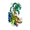
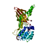
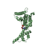
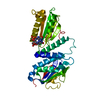
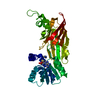
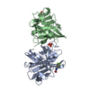
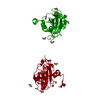
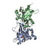
- PDB-3cwv: Crystal structure of B-subunit of the DNA gyrase from Myxococcus ... -
+
Open data
ID or keywords:
Loading...
-
Basic information
Entry
Database: PDB / ID: 3cwv
Title
Crystal structure of B-subunit of the DNA gyrase from Myxococcus xanthus
Components
DNA gyrase, B subunit, truncated
Keywords
ISOMERASE / structural genomics / UNKNOWN FUNCTION / DNA Gyrase / B-Subunit / ATP-binding / Nucleotide-binding / Topoisomerase / PSI-2 / Protein Structure Initiative / New York SGX Research Center for Structural Genomics / NYSGXRC
Function / homology
Function and homology information
DNA topoisomerase type II (double strand cut, ATP-hydrolyzing) activity / DNA topoisomerase (ATP-hydrolysing) / DNA topological change / DNA binding / ATP binding Similarity search - Function
DNA topoisomerase, type IIA, subunit B / DNA topoisomerase, type IIA, subunit B, domain 2 / DNA gyrase B / Ribosomal Protein S5; domain 2 - #10 / Ribosomal Protein S5; domain 2 / Histidine kinase-like ATPase, C-terminal domain / Heat Shock Protein 90 / Histidine kinase/HSP90-like ATPase superfamily / Ribosomal protein S5 domain 2-type fold, subgroup / Ribosomal protein S5 domain 2-type fold ...DNA topoisomerase, type IIA, subunit B / DNA topoisomerase, type IIA, subunit B, domain 2 / DNA gyrase B / Ribosomal Protein S5; domain 2 - #10 / Ribosomal Protein S5; domain 2 / Histidine kinase-like ATPase, C-terminal domain / Heat Shock Protein 90 / Histidine kinase/HSP90-like ATPase superfamily / Ribosomal protein S5 domain 2-type fold, subgroup / Ribosomal protein S5 domain 2-type fold / 2-Layer Sandwich / Alpha Beta Similarity search - Domain/homology
Protocol: SINGLE WAVELENGTH / Monochromatic (M) / Laue (L): M / Scattering type: x-ray
Radiation wavelength
Wavelength: 0.9793 Å / Relative weight: 1
Reflection
Redundancy: 2.5 % / Av σ(I) over netI: 11.7 / Number: 302348 / Rmerge(I) obs: 0.048 / Χ2: 0.8 / D res high: 1.95 Å / D res low: 50 Å / Num. obs: 120809 / % possible obs: 96.5
In the structure databanks used in Yorodumi, some data are registered as the other names, "COVID-19 virus" and "2019-nCoV". Here are the details of the virus and the list of structure data.
Jan 31, 2019. EMDB accession codes are about to change! (news from PDBe EMDB page)
EMDB accession codes are about to change! (news from PDBe EMDB page)
The allocation of 4 digits for EMDB accession codes will soon come to an end. Whilst these codes will remain in use, new EMDB accession codes will include an additional digit and will expand incrementally as the available range of codes is exhausted. The current 4-digit format prefixed with “EMD-” (i.e. EMD-XXXX) will advance to a 5-digit format (i.e. EMD-XXXXX), and so on. It is currently estimated that the 4-digit codes will be depleted around Spring 2019, at which point the 5-digit format will come into force.
The EM Navigator/Yorodumi systems omit the EMD- prefix.
Related info.:Q: What is EMD? / ID/Accession-code notation in Yorodumi/EM Navigator
Yorodumi is a browser for structure data from EMDB, PDB, SASBDB, etc.
This page is also the successor to EM Navigator detail page, and also detail information page/front-end page for Omokage search.
The word "yorodu" (or yorozu) is an old Japanese word meaning "ten thousand". "mi" (miru) is to see.
Related info.:EMDB / PDB / SASBDB / Comparison of 3 databanks / Yorodumi Search / Aug 31, 2016. New EM Navigator & Yorodumi / Yorodumi Papers / Jmol/JSmol / Function and homology information / Changes in new EM Navigator and Yorodumi
 Movie
Movie Controller

 Yorodumi
 Open data
 Basic information
 Components
 Keywords
 Function and homology information
 Myxococcus xanthus (bacteria)
 X-RAY DIFFRACTION /
  Authors
 Citation
 Structure visualization
 Downloads & links
 Other downloads
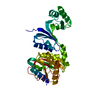
 PDBj
PDBj

 Assembly


 Mass: 18.015 Da / Num. of mol.: 345 / Source method: isolated from a natural source / Formula: H2O
 Sample preparation
 / Beamline: 24-ID-C / Wavelength: 0.9793 Å
 Processing