+ Open data
- Basic information
| Entry | Database: PDB / ID: 7ppj | |||||||||||||||||||||||||||||||||||||||||||||
|---|---|---|---|---|---|---|---|---|---|---|---|---|---|---|---|---|---|---|---|---|---|---|---|---|---|---|---|---|---|---|---|---|---|---|---|---|---|---|---|---|---|---|---|---|---|---|
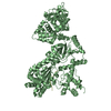
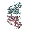
| Title | human SLFN5 | |||||||||||||||||||||||||||||||||||||||||||||
 Components Components | Schlafen family member 5 | |||||||||||||||||||||||||||||||||||||||||||||
 Keywords Keywords | DNA BINDING PROTEIN / nucleotide binding protein antiviral linked to tumorigenesis transcription regulation | |||||||||||||||||||||||||||||||||||||||||||||
| Function / homology |  Function and homology information Function and homology information | |||||||||||||||||||||||||||||||||||||||||||||
| Biological species |  Homo sapiens (human) Homo sapiens (human) | |||||||||||||||||||||||||||||||||||||||||||||
| Method | ELECTRON MICROSCOPY / single particle reconstruction / cryo EM / Resolution: 3.44 Å | |||||||||||||||||||||||||||||||||||||||||||||
 Authors Authors | Lammens, K. / Metzner, F.J. | |||||||||||||||||||||||||||||||||||||||||||||
| Funding support |  Germany, 1items Germany, 1items
| |||||||||||||||||||||||||||||||||||||||||||||
 Citation Citation |  Journal: Nucleic Acids Res / Year: 2022 Journal: Nucleic Acids Res / Year: 2022Title: Structural and biochemical characterization of human Schlafen 5. Authors: Felix J Metzner / Elisabeth Huber / Karl-Peter Hopfner / Katja Lammens /  Abstract: The Schlafen family belongs to the interferon-stimulated genes and its members are involved in cell cycle regulation, T cell quiescence, inhibition of viral replication, DNA-repair and tRNA ...The Schlafen family belongs to the interferon-stimulated genes and its members are involved in cell cycle regulation, T cell quiescence, inhibition of viral replication, DNA-repair and tRNA processing. Here, we present the cryo-EM structure of full-length human Schlafen 5 (SLFN5) and the high-resolution crystal structure of the highly conserved N-terminal core domain. We show that the core domain does not resemble an ATPase-like fold and neither binds nor hydrolyzes ATP. SLFN5 binds tRNA as well as single- and double-stranded DNA, suggesting a potential role in transcriptional regulation. Unlike rat Slfn13 or human SLFN11, human SLFN5 did not cleave tRNA. Based on the structure, we identified two residues in proximity to the zinc finger motif that decreased DNA binding when mutated. These results indicate that Schlafen proteins have divergent enzymatic functions and provide a structural platform for future biochemical and genetic studies. | |||||||||||||||||||||||||||||||||||||||||||||
| History |
|
- Structure visualization
| Movie |
 Movie viewer Movie viewer |
|---|---|
| Structure viewer | Molecule:  Molmil Molmil Jmol/JSmol Jmol/JSmol |
- Downloads & links
- Download
| PDBx/mmCIF format |  7ppj.cif.gz 7ppj.cif.gz | 132.6 KB | Display |  PDBx/mmCIF format PDBx/mmCIF format |
|---|---|---|---|---|
| PDB format |  pdb7ppj.ent.gz pdb7ppj.ent.gz | 97.3 KB | Display |  PDB format PDB format |
| PDBx/mmJSON format |  7ppj.json.gz 7ppj.json.gz | Tree view |  PDBx/mmJSON format PDBx/mmJSON format | |
| Others |  Other downloads Other downloads |
-Validation report
| Summary document |  7ppj_validation.pdf.gz 7ppj_validation.pdf.gz | 1.1 MB | Display |  wwPDB validaton report wwPDB validaton report |
|---|---|---|---|---|
| Full document |  7ppj_full_validation.pdf.gz 7ppj_full_validation.pdf.gz | 1.1 MB | Display | |
| Data in XML |  7ppj_validation.xml.gz 7ppj_validation.xml.gz | 31.6 KB | Display | |
| Data in CIF |  7ppj_validation.cif.gz 7ppj_validation.cif.gz | 44.6 KB | Display | |
| Arichive directory |  https://data.pdbj.org/pub/pdb/validation_reports/pp/7ppj https://data.pdbj.org/pub/pdb/validation_reports/pp/7ppj ftp://data.pdbj.org/pub/pdb/validation_reports/pp/7ppj ftp://data.pdbj.org/pub/pdb/validation_reports/pp/7ppj | HTTPS FTP |
-Related structure data
| Related structure data |  13581MC 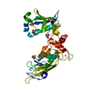 6rr9C  7q3zC C: citing same article ( M: map data used to model this data |
|---|---|
| Similar structure data |
- Links
Links
- Assembly
| Deposited unit | 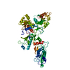
|
|---|---|
| 1 |
|
- Components
| #1: Protein | Mass: 104420.016 Da / Num. of mol.: 1 Source method: isolated from a genetically manipulated source Details: N-terminal 2 x Flag tag HRV 3C site flexible C-terminus Source: (gene. exp.)  Homo sapiens (human) / Gene: SLFN5 / Production host: Homo sapiens (human) / Gene: SLFN5 / Production host:  Homo sapiens (human) / References: UniProt: Q08AF3 Homo sapiens (human) / References: UniProt: Q08AF3 |
|---|---|
| #2: Chemical | ChemComp-ZN / |
| Has ligand of interest | N |
| Has protein modification | N |
-Experimental details
-Experiment
| Experiment | Method: ELECTRON MICROSCOPY |
|---|---|
| EM experiment | Aggregation state: PARTICLE / 3D reconstruction method: single particle reconstruction |
- Sample preparation
| Component | Name: full length human Schlafen 5 / Type: ORGANELLE OR CELLULAR COMPONENT / Entity ID: #1 / Source: RECOMBINANT |
|---|---|
| Molecular weight | Experimental value: NO |
| Source (natural) | Organism:  Homo sapiens (human) Homo sapiens (human) |
| Source (recombinant) | Organism:  Homo sapiens (human) / Cell: Expi293F / Plasmid: pcDNA3.1 Homo sapiens (human) / Cell: Expi293F / Plasmid: pcDNA3.1 |
| Buffer solution | pH: 9 |
| Specimen | Embedding applied: NO / Shadowing applied: NO / Staining applied: NO / Vitrification applied: YES |
| Vitrification | Cryogen name: ETHANE |
- Electron microscopy imaging
| Experimental equipment |  Model: Titan Krios / Image courtesy: FEI Company |
|---|---|
| Microscopy | Model: FEI TITAN KRIOS |
| Electron gun | Electron source:  FIELD EMISSION GUN / Accelerating voltage: 300 kV / Illumination mode: FLOOD BEAM FIELD EMISSION GUN / Accelerating voltage: 300 kV / Illumination mode: FLOOD BEAM |
| Electron lens | Mode: BRIGHT FIELD |
| Image recording | Electron dose: 41 e/Å2 / Film or detector model: GATAN K2 SUMMIT (4k x 4k) |
- Processing
| Software | Name: PHENIX / Version: 1.17.1_3660: / Classification: refinement | ||||||||||||||||||||||||
|---|---|---|---|---|---|---|---|---|---|---|---|---|---|---|---|---|---|---|---|---|---|---|---|---|---|
| EM software | Name: PHENIX / Category: model refinement | ||||||||||||||||||||||||
| CTF correction | Type: PHASE FLIPPING AND AMPLITUDE CORRECTION | ||||||||||||||||||||||||
| 3D reconstruction | Resolution: 3.44 Å / Resolution method: FSC 0.143 CUT-OFF / Num. of particles: 140715 / Symmetry type: POINT | ||||||||||||||||||||||||
| Refine LS restraints |
|
 Movie
Movie Controller



 PDBj
PDBj

