 Movie
Movie Controller


+ Open data

- Basic information
| Entry | Database: PDB / ID: 7pmx | ||||||
|---|---|---|---|---|---|---|---|
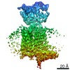
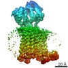
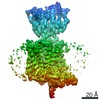
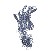
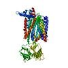
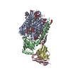
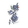
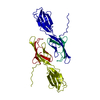
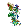
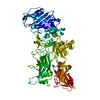
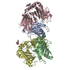
| Title | HsPepT1 bound to Ala-Phe in the outward facing open conformation | ||||||
 Components Components |
| ||||||
 Keywords Keywords |  MEMBRANE PROTEIN / HsPepT1 / PepT1 / Peptide transporter MEMBRANE PROTEIN / HsPepT1 / PepT1 / Peptide transporter | ||||||
| Function / homology |  Function and homology information Function and homology informationproton-dependent oligopeptide secondary active transmembrane transporter activity / tripeptide import across plasma membrane / Proton/oligopeptide cotransporters / tripeptide transmembrane transporter activity / dipeptide import across plasma membrane / peptide:proton symporter activity / dipeptide transmembrane transporter activity / brush border / monoatomic ion transport / protein transport ...proton-dependent oligopeptide secondary active transmembrane transporter activity / tripeptide import across plasma membrane / Proton/oligopeptide cotransporters / tripeptide transmembrane transporter activity / dipeptide import across plasma membrane / peptide:proton symporter activity / dipeptide transmembrane transporter activity / brush border / monoatomic ion transport / protein transport / apical plasma membrane / membrane / plasma membraneSimilarity search - Function | ||||||
| Biological species |  Homo sapiens (human) Homo sapiens (human) | ||||||
| Method | ELECTRON MICROSCOPY / single particle reconstruction / cryo EM / Resolution: 3.5 Å | ||||||
 Authors Authors | Killer, M. / Wald, J. / Pieprzyk, J. / Marlovits, T.C. / Loew, C. | ||||||
| Funding support | 1items
| ||||||
 Citation Citation | Journal: Sci Adv / Year: 2021 Title: Structural snapshots of human PepT1 and PepT2 reveal mechanistic insights into substrate and drug transport across epithelial membranes. Authors: Maxime Killer / Jiri Wald / Joanna Pieprzyk / Thomas C Marlovits / Christian Löw /  Abstract: The uptake of peptides in mammals plays a crucial role in nutrition and inflammatory diseases. This process is mediated by promiscuous transporters of the solute carrier family 15, which form part of ...The uptake of peptides in mammals plays a crucial role in nutrition and inflammatory diseases. This process is mediated by promiscuous transporters of the solute carrier family 15, which form part of the major facilitator superfamily. Besides the uptake of short peptides, peptide transporter 1 (PepT1) is a highly abundant drug transporter in the intestine and represents a major route for oral drug delivery. PepT2 also allows renal drug reabsorption from ultrafiltration and brain-to-blood efflux of neurotoxic compounds. Here, we present cryogenic electron microscopy (cryo-EM) structures of human PepT1 and PepT2 captured in four different states throughout the transport cycle. The structures reveal the architecture of human peptide transporters and provide mechanistic insights into substrate recognition and conformational transitions during transport. This may support future drug design efforts to increase the bioavailability of different drugs in the human body. | ||||||
| History |
|
- Structure visualization
| Movie |
Movie viewer |
|---|---|
| Structure viewer | Molecule: MolmilJmol/JSmol |
- Downloads & links
-Download
| PDBx/mmCIF format | 7pmx.cif.gz | 220.6 KB | Display | PDBx/mmCIF format |
|---|---|---|---|---|
| PDB format | pdb7pmx.ent.gz | 185.4 KB | Display | PDB format |
| PDBx/mmJSON format | 7pmx.json.gz | Tree view | PDBx/mmJSON format | |
| Others |  Other downloads Other downloads |
-Validation report
| Arichive directory | https://data.pdbj.org/pub/pdb/validation_reports/pm/7pmxftp://data.pdbj.org/pub/pdb/validation_reports/pm/7pmx | HTTPS FTP |
|---|
-Related structure data
| Related structure data | 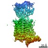 13543MC 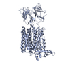 7pmwC 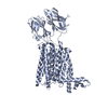 7pmyC 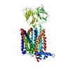 7pn1C M: map data used to model this data C: citing same article ( |
|---|---|
| Similar structure data |
-Links
 PDBj
PDBj


- Assembly
| Deposited unit | 
|
|---|---|
| 1 |
|
-Components
| #1: Protein | Mass: 78872.422 Da / Num. of mol.: 1 Source method: isolated from a genetically manipulated source Details: HsPepT1 / Source: (gene. exp.) Homo sapiens (human) / Gene: SLC15A1, PEPT1 / Production host: Homo sapiens (human) / References: UniProt: P46059 |
|---|---|
| #2: Protein/peptide | Mass: 236.267 Da / Num. of mol.: 1 / Source method: obtained synthetically / Source: (synth.) Homo sapiens (human) |
-Experimental details
-Experiment
| Experiment | Method: ELECTRON MICROSCOPY |
|---|---|
| EM experiment | Aggregation state: PARTICLE / 3D reconstruction method: single particle reconstruction |
- Sample preparation
| Component |
| ||||||||||||||||||
|---|---|---|---|---|---|---|---|---|---|---|---|---|---|---|---|---|---|---|---|
| Molecular weight | Experimental value: NO | ||||||||||||||||||
| Source (natural) |
| ||||||||||||||||||
| Source (recombinant) |
| ||||||||||||||||||
| Buffer solution | pH: 7.5 | ||||||||||||||||||
| Specimen | Conc.: 2 mg/ml / Embedding applied: NO / Shadowing applied: NO / Staining applied: NO / Vitrification applied: YES | ||||||||||||||||||
| Vitrification | Instrument: FEI VITROBOT MARK IV / Cryogen name: PROPANE / Humidity: 100 % / Chamber temperature: 278 K |
- Electron microscopy imaging
| Experimental equipment |  Model: Titan Krios / Image courtesy: FEI Company |
|---|---|
| Microscopy | Model: FEI TITAN KRIOS |
| Electron gun | Electron source: FIELD EMISSION GUN / Accelerating voltage: 300 kV / Illumination mode: FLOOD BEAM |
| Electron lens | Mode: BRIGHT FIELDBright-field microscopy |
| Image recording | Electron dose: 55 e/Å2 / Film or detector model: GATAN K3 BIOQUANTUM (6k x 4k) |
- Processing
| EM software | Name: cryoSPARC / Version: 3 / Category: final Euler assignment |
|---|---|
| CTF correction | Type: PHASE FLIPPING AND AMPLITUDE CORRECTION |
| 3D reconstruction | Resolution: 3.5 Å / Resolution method: FSC 0.143 CUT-OFF / Num. of particles: 466042 / Symmetry type: POINT |
