 Movie
Movie Controller

[English] 日本語
 Yorodumi
- PDB-3ehb: A D-Pathway Mutation Decouples the Paracoccus Denitrificans Cytoc... -
+ Open data

- Basic information
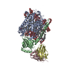
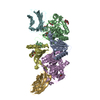
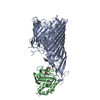
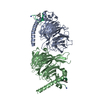
| Entry | Database: PDB / ID: 3ehb | ||||||
|---|---|---|---|---|---|---|---|
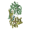
| Title | A D-Pathway Mutation Decouples the Paracoccus Denitrificans Cytochrome c Oxidase by Altering the side chain orientation of a distant, conserved Glutamate | ||||||
 Components Components |
| ||||||
 Keywords Keywords | OXIDOREDUCTASE/IMMUNE SYSTEM / proton pumping / water chain / electron transfer / Cell inner membrane / Cell membrane / Copper / Electron transport / Heme / Hydrogen ion transport / Ion transport / Iron / Membrane / Metal-binding / Oxidoreductase / Respiratory chain / Transmembrane / Transport / Pyrrolidone carboxylic acid / OXIDOREDUCTASE-IMMUNE SYSTEM COMPLEX | ||||||
| Function / homology |  Function and homology information Function and homology informationrespiratory chain complex IV / cytochrome-c oxidase / oxidative phosphorylation / cytochrome-c oxidase activity / immunoglobulin mediated immune response / electron transport coupled proton transport / immunoglobulin complex / antigen binding / ATP synthesis coupled electron transport / respiratory electron transport chain ...respiratory chain complex IV / cytochrome-c oxidase / oxidative phosphorylation / cytochrome-c oxidase activity / immunoglobulin mediated immune response / electron transport coupled proton transport / immunoglobulin complex / antigen binding / ATP synthesis coupled electron transport / respiratory electron transport chain / adaptive immune response / oxidoreductase activity / immune response / copper ion binding / heme binding / extracellular region / metal ion binding / plasma membrane Similarity search - Function | ||||||
| Biological species |  Paracoccus denitrificans (bacteria) Paracoccus denitrificans (bacteria) | ||||||
| Method |  X-RAY DIFFRACTION / SYNCHROTRON / MOLECULAR REPLACEMENT / Resolution: 2.32 Å X-RAY DIFFRACTION / SYNCHROTRON / MOLECULAR REPLACEMENT / Resolution: 2.32 Å | ||||||
 Authors Authors | Koepke, J. / Mueller, H. / Peng, G. | ||||||
 Citation Citation | Journal: J.Mol.Biol. / Year: 2008 Title: A d-pathway mutation decouples the paracoccusdenitrificans cytochrome C oxidase by altering the side-chain orientation of a distant conserved glutamate. Authors: Durr, K.L. / Koepke, J. / Hellwig, P. / Muller, H. / Angerer, H. / Peng, G. / Olkhova, E. / Richter, O.-M.H. / Ludwig, B. / Michel, H. | ||||||
| History |
|
- Structure visualization
| Structure viewer | Molecule: MolmilJmol/JSmol |
|---|
- Downloads & links
-Download
| PDBx/mmCIF format | 3ehb.cif.gz | 246.6 KB | Display | PDBx/mmCIF format |
|---|---|---|---|---|
| PDB format | pdb3ehb.ent.gz | 194.1 KB | Display | PDB format |
| PDBx/mmJSON format | 3ehb.json.gz | Tree view | PDBx/mmJSON format | |
| Others |  Other downloads Other downloads |
-Validation report
| Arichive directory | https://data.pdbj.org/pub/pdb/validation_reports/eh/3ehbftp://data.pdbj.org/pub/pdb/validation_reports/eh/3ehb | HTTPS FTP |
|---|
-Related structure data
| Related structure data |  1ar1S S: Starting model for refinement |
|---|---|
| Similar structure data |


-Links
 PDBj
PDBj


- Assembly
| Deposited unit | 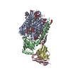
| ||||||||
|---|---|---|---|---|---|---|---|---|---|
| 1 |
| ||||||||
| Unit cell |
|
-Components
-Cytochrome c oxidase subunit ... , 2 types, 2 molecules AB
| #1: Protein | Mass: 62487.629 Da / Num. of mol.: 1 / Fragment: UNP residues 17-545 / Mutation: N131D Source method: isolated from a genetically manipulated source Source: (gene. exp.) Paracoccus denitrificans (bacteria) / Production host: |
|---|---|
| #2: Protein | Mass: 32563.643 Da / Num. of mol.: 1 Source method: isolated from a genetically manipulated source Source: (gene. exp.) Paracoccus denitrificans (bacteria) / Production host: |
-Antibody , 2 types, 2 molecules CD
| #3: Antibody | Mass: 14324.923 Da / Num. of mol.: 1 Source method: isolated from a genetically manipulated source Source: (gene. exp.) |
|---|---|
| #4: Antibody | Mass: 13260.795 Da / Num. of mol.: 1 Source method: isolated from a genetically manipulated source Source: (gene. exp.) |
-Sugars , 1 types, 12 molecules 
| #10: Sugar | ChemComp-LMT /  Type: D-saccharide / Mass: 510.615 Da / Num. of mol.: 12 / Source method: obtained synthetically / Formula: C24H46O11 / Comment: detergent*YM Type: D-saccharide / Mass: 510.615 Da / Num. of mol.: 12 / Source method: obtained synthetically / Formula: C24H46O11 / Comment: detergent*YM |
|---|
-Non-polymers , 7 types, 396 molecules 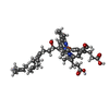


| #5: Chemical |  Mass: 852.837 Da / Num. of mol.: 2 / Source method: obtained synthetically / Formula: C49H56FeN4O6 Mass: 852.837 Da / Num. of mol.: 2 / Source method: obtained synthetically / Formula: C49H56FeN4O6#6: Chemical |  Mass: 63.546 Da / Num. of mol.: 3 / Source method: obtained synthetically / Formula: Cu Mass: 63.546 Da / Num. of mol.: 3 / Source method: obtained synthetically / Formula: Cu#7: Chemical | ChemComp-MG / |  Mass: 24.305 Da / Num. of mol.: 1 / Source method: obtained synthetically / Formula: Mg Mass: 24.305 Da / Num. of mol.: 1 / Source method: obtained synthetically / Formula: Mg#8: Chemical | ChemComp-CA / |  Mass: 40.078 Da / Num. of mol.: 1 / Source method: obtained synthetically / Formula: Ca Mass: 40.078 Da / Num. of mol.: 1 / Source method: obtained synthetically / Formula: Ca#9: Chemical | ChemComp-LDA /  Mass: 229.402 Da / Num. of mol.: 9 / Source method: obtained synthetically / Formula: C14H31NO / Comment: LDAO, detergent*YM Mass: 229.402 Da / Num. of mol.: 9 / Source method: obtained synthetically / Formula: C14H31NO / Comment: LDAO, detergent*YM#11: Chemical | ChemComp-PER / |  Mass: 31.999 Da / Num. of mol.: 1 / Source method: obtained synthetically / Formula: O2 Mass: 31.999 Da / Num. of mol.: 1 / Source method: obtained synthetically / Formula: O2#12: Water | ChemComp-HOH / | Mass: 18.015 Da / Num. of mol.: 379 / Source method: isolated from a natural source / Formula: H2O |
|---|
-Details
| Has protein modification | Y |
|---|
-Experimental details
-Experiment
| Experiment | Method: X-RAY DIFFRACTION / Number of used crystals: 1 |
|---|
- Sample preparation
| Crystal | Density Matthews: 4.1 Å3/Da / Density % sol: 70.03 % Description: 25% (v/v) glycerol was used as cryoprotectant to flash freeze in a gaseous liquid nitrogen cooled stream |
|---|---|
| Crystal grow | Temperature: 291 K / Method: vapor diffusion, hanging drop / pH: 7.3 Details: MPEG 2000 is contained in the crystallization buffer pH 5.5, pH 7.3, VAPOR DIFFUSION, HANGING DROP, temperature 291K |
-Data collection
| Diffraction | Mean temperature: 100 K |
|---|---|
| Diffraction source | Source: SYNCHROTRON / Site: SLS  / Beamline: X10SA / Wavelength: 0.92021 Å / Beamline: X10SA / Wavelength: 0.92021 Å |
| Detector | Type: MARMOSAIC 225 mm CCD / Detector: CCD / Date: Nov 16, 2006 |
| Radiation | Monochromator: double mirror / Protocol: SINGLE WAVELENGTH / Monochromatic (M) / Laue (L): M / Scattering type: x-ray |
| Radiation wavelength | Wavelength: 0.92021 Å / Relative weight: 1 |
| Reflection | Resolution: 2.32→20 Å / Num. all: 87146 / Num. obs: 87146 / % possible obs: 98.8 % / Observed criterion σ(F): -3 / Observed criterion σ(I): -3 / Biso Wilson estimate: 51.5 Å2 / Rmerge(I) obs: 0.09 / Net I/σ(I): 19.14 |
| Reflection shell | Resolution: 2.32→2.4 Å / Mean I/σ(I) obs: 3.93 / Num. unique all: 8161 / % possible all: 97 |
- Processing
| Software |
| ||||||||||||||||||||||||||||||||||||||||||||||||||||||||||||||||||||||||||||||||||||||||||||||||||||||||||||||||||||||||||||||||||||||||||||||||||||||||||||||||||||||||||
|---|---|---|---|---|---|---|---|---|---|---|---|---|---|---|---|---|---|---|---|---|---|---|---|---|---|---|---|---|---|---|---|---|---|---|---|---|---|---|---|---|---|---|---|---|---|---|---|---|---|---|---|---|---|---|---|---|---|---|---|---|---|---|---|---|---|---|---|---|---|---|---|---|---|---|---|---|---|---|---|---|---|---|---|---|---|---|---|---|---|---|---|---|---|---|---|---|---|---|---|---|---|---|---|---|---|---|---|---|---|---|---|---|---|---|---|---|---|---|---|---|---|---|---|---|---|---|---|---|---|---|---|---|---|---|---|---|---|---|---|---|---|---|---|---|---|---|---|---|---|---|---|---|---|---|---|---|---|---|---|---|---|---|---|---|---|---|---|---|---|---|---|
| Refinement | Method to determine structure: MOLECULAR REPLACEMENT Starting model: PDB ENTRY 1AR1 Resolution: 2.32→19.69 Å / Cor.coef. Fo:Fc: 0.932 / Cor.coef. Fo:Fc free: 0.912 / SU B: 5.594 / SU ML: 0.14 / Cross valid method: THROUGHOUT / σ(F): -3 / ESU R: 0.208 / ESU R Free: 0.186 / Stereochemistry target values: MAXIMUM LIKELIHOOD Details: Due to a feature in the refinement program, the structure was refined with OXT on one or more residues that are not the terminal residues of the sequence. In all these instances the OXT was ...Details: Due to a feature in the refinement program, the structure was refined with OXT on one or more residues that are not the terminal residues of the sequence. In all these instances the OXT was changed to N of the next residue
| ||||||||||||||||||||||||||||||||||||||||||||||||||||||||||||||||||||||||||||||||||||||||||||||||||||||||||||||||||||||||||||||||||||||||||||||||||||||||||||||||||||||||||
| Solvent computation | Ion probe radii: 0.8 Å / Shrinkage radii: 0.8 Å / VDW probe radii: 1.2 Å / Solvent model: BABINET MODEL WITH MASK | ||||||||||||||||||||||||||||||||||||||||||||||||||||||||||||||||||||||||||||||||||||||||||||||||||||||||||||||||||||||||||||||||||||||||||||||||||||||||||||||||||||||||||
| Displacement parameters | Biso mean: 60.153 Å2 | ||||||||||||||||||||||||||||||||||||||||||||||||||||||||||||||||||||||||||||||||||||||||||||||||||||||||||||||||||||||||||||||||||||||||||||||||||||||||||||||||||||||||||
| Refinement step | Cycle: LAST / Resolution: 2.32→19.69 Å
| ||||||||||||||||||||||||||||||||||||||||||||||||||||||||||||||||||||||||||||||||||||||||||||||||||||||||||||||||||||||||||||||||||||||||||||||||||||||||||||||||||||||||||
| Refine LS restraints |
| ||||||||||||||||||||||||||||||||||||||||||||||||||||||||||||||||||||||||||||||||||||||||||||||||||||||||||||||||||||||||||||||||||||||||||||||||||||||||||||||||||||||||||
| LS refinement shell | Resolution: 2.318→2.377 Å / Total num. of bins used: 20
|
