 Movie
Movie Controller

[English] 日本語
 Yorodumi
- PDB-7pj4: Crystal structure of catalytic domain in closed conformation of L... -
+ Open data

- Basic information
| Entry | Database: PDB / ID: 7pj4 | ||||||
|---|---|---|---|---|---|---|---|
| Title | Crystal structure of catalytic domain in closed conformation of LytB (E585Q)from Streptococcus pneumoniae | ||||||
 Components Components | Putative endo-beta-N-acetylglucosaminidase | ||||||
 Keywords Keywords | HYDROLASE / glucosaminidase / peptidoglycan hydrolase | ||||||
| Function / homology |  Function and homology information Function and homology informationmannosyl-glycoprotein endo-beta-N-acetylglucosaminidase / mannosyl-glycoprotein endo-beta-N-acetylglucosaminidase activity / amidase activity / cell wall organization / extracellular region Similarity search - Function | ||||||
| Biological species |   Streptococcus pneumoniae (bacteria) Streptococcus pneumoniae (bacteria) | ||||||
| Method |  X-RAY DIFFRACTION / SYNCHROTRON / MOLECULAR REPLACEMENT / Resolution: 1.25 Å X-RAY DIFFRACTION / SYNCHROTRON / MOLECULAR REPLACEMENT / Resolution: 1.25 Å | ||||||
 Authors Authors | Martinez Caballero, S. / Hermoso, J.A. | ||||||
| Funding support | 1items
| ||||||
 Citation Citation | Journal: Cell Rep / Year: 2023 Title: Molecular basis of the final step of cell division in Streptococcus pneumoniae. Authors: Martinez-Caballero, S. / Freton, C. / Molina, R. / Bartual, S.G. / Gueguen-Chaignon, V. / Mercy, C. / Gago, F. / Mahasenan, K.V. / Munoz, I.G. / Lee, M. / Hesek, D. / Mobashery, S. / ...Authors: Martinez-Caballero, S. / Freton, C. / Molina, R. / Bartual, S.G. / Gueguen-Chaignon, V. / Mercy, C. / Gago, F. / Mahasenan, K.V. / Munoz, I.G. / Lee, M. / Hesek, D. / Mobashery, S. / Hermoso, J.A. / Grangeasse, C. | ||||||
| History |
|
- Structure visualization
| Structure viewer | Molecule: MolmilJmol/JSmol |
|---|
- Downloads & links
-Download
| PDBx/mmCIF format | 7pj4.cif.gz | 134.8 KB | Display | PDBx/mmCIF format |
|---|---|---|---|---|
| PDB format | pdb7pj4.ent.gz | Display | PDB format | |
| PDBx/mmJSON format | 7pj4.json.gz | Tree view | PDBx/mmJSON format | |
| Others |  Other downloads Other downloads |
-Validation report
| Arichive directory | https://data.pdbj.org/pub/pdb/validation_reports/pj/7pj4ftp://data.pdbj.org/pub/pdb/validation_reports/pj/7pj4 | HTTPS FTP |
|---|
-Related structure data
| Related structure data | 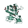 7pj3SC 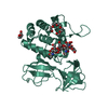 7pj5C  7pj6C  7pl2C  7pl3C  7pl5C  7podC S: Starting model for refinement C: citing same article ( |
|---|---|
| Similar structure data |
-Links
 PDBj
PDBj
- Assembly
| Deposited unit | 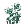
| ||||||||
|---|---|---|---|---|---|---|---|---|---|
| 1 |
| ||||||||
| Unit cell |
|
-Components
-Protein , 1 types, 1 molecules AAA
| #1: Protein | Mass: 30895.436 Da / Num. of mol.: 1 / Mutation: E585Q Source method: isolated from a genetically manipulated source Source: (gene. exp.) Streptococcus pneumoniae (strain ATCC BAA-255 / R6) (bacteria)Strain: ATCC BAA-255 / R6 / Gene: lytB, spr0867 / Production host: References: UniProt: P59206, mannosyl-glycoprotein endo-beta-N-acetylglucosaminidase |
|---|
-Non-polymers , 6 types, 306 molecules 


| #2: Chemical | ChemComp-1PE /  Mass: 238.278 Da / Num. of mol.: 1 / Source method: obtained synthetically / Formula: C10H22O6 / Comment: precipitant*YM Mass: 238.278 Da / Num. of mol.: 1 / Source method: obtained synthetically / Formula: C10H22O6 / Comment: precipitant*YM |
|---|---|
| #3: Chemical | ChemComp-PGE /  Mass: 150.173 Da / Num. of mol.: 1 / Source method: obtained synthetically / Formula: C6H14O4 Mass: 150.173 Da / Num. of mol.: 1 / Source method: obtained synthetically / Formula: C6H14O4 |
| #4: Chemical | ChemComp-PEG /  Mass: 106.120 Da / Num. of mol.: 1 / Source method: obtained synthetically / Formula: C4H10O3 Mass: 106.120 Da / Num. of mol.: 1 / Source method: obtained synthetically / Formula: C4H10O3 |
| #5: Chemical | ChemComp-ACT /  Mass: 59.044 Da / Num. of mol.: 1 / Source method: obtained synthetically / Formula: C2H3O2 Mass: 59.044 Da / Num. of mol.: 1 / Source method: obtained synthetically / Formula: C2H3O2 |
| #6: Chemical | ChemComp-CA /  Mass: 40.078 Da / Num. of mol.: 1 / Source method: obtained synthetically / Formula: Ca Mass: 40.078 Da / Num. of mol.: 1 / Source method: obtained synthetically / Formula: Ca |
| #7: Water | ChemComp-HOH / Mass: 18.015 Da / Num. of mol.: 301 / Source method: isolated from a natural source / Formula: H2O |
-Details
| Has ligand of interest | N |
|---|
-Experimental details
-Experiment
| Experiment | Method: X-RAY DIFFRACTION / Number of used crystals: 1 |
|---|
- Sample preparation
| Crystal | Density Matthews: 2.17 Å3/Da / Density % sol: 43.38 % |
|---|---|
| Crystal grow | Temperature: 291.15 K / Method: vapor diffusion, sitting drop / Details: PEG 300 Bis-Tris buffer Calcium acetate |
-Data collection
| Diffraction | Mean temperature: 100 K / Serial crystal experiment: N |
|---|---|
| Diffraction source | Source: SYNCHROTRON / Site: ALBA  / Beamline: XALOC / Wavelength: 0.97926 Å / Beamline: XALOC / Wavelength: 0.97926 Å |
| Detector | Type: DECTRIS PILATUS 6M / Detector: PIXEL / Date: Nov 8, 2019 |
| Radiation | Protocol: SINGLE WAVELENGTH / Monochromatic (M) / Laue (L): M / Scattering type: x-ray |
| Radiation wavelength | Wavelength: 0.97926 Å / Relative weight: 1 |
| Reflection | Resolution: 1.25→46.34 Å / Num. obs: 74735 / % possible obs: 100 % / Redundancy: 12 % / Biso Wilson estimate: 12.1 Å2 / Rmerge(I) obs: 0.066 / Rpim(I) all: 0.02 / Net I/σ(I): 20.4 |
| Reflection shell | Resolution: 1.25→1.27 Å / Rmerge(I) obs: 0.763 / Num. unique obs: 3690 / Rpim(I) all: 0.226 |
- Processing
| Software |
| |||||||||||||||||||||||||||||||||||||||||||||||||||||||||||||||||||||||||||||||||||||||||||||||||||||||||||||||||||||||||||||||||||||||||||||||||||||||||||||||||||||||||||||||||||||||||||||||||||||||||||||||||||||||||||||||||||||||
|---|---|---|---|---|---|---|---|---|---|---|---|---|---|---|---|---|---|---|---|---|---|---|---|---|---|---|---|---|---|---|---|---|---|---|---|---|---|---|---|---|---|---|---|---|---|---|---|---|---|---|---|---|---|---|---|---|---|---|---|---|---|---|---|---|---|---|---|---|---|---|---|---|---|---|---|---|---|---|---|---|---|---|---|---|---|---|---|---|---|---|---|---|---|---|---|---|---|---|---|---|---|---|---|---|---|---|---|---|---|---|---|---|---|---|---|---|---|---|---|---|---|---|---|---|---|---|---|---|---|---|---|---|---|---|---|---|---|---|---|---|---|---|---|---|---|---|---|---|---|---|---|---|---|---|---|---|---|---|---|---|---|---|---|---|---|---|---|---|---|---|---|---|---|---|---|---|---|---|---|---|---|---|---|---|---|---|---|---|---|---|---|---|---|---|---|---|---|---|---|---|---|---|---|---|---|---|---|---|---|---|---|---|---|---|---|---|---|---|---|---|---|---|---|---|---|---|---|---|---|---|---|---|
| Refinement | Method to determine structure: MOLECULAR REPLACEMENT Starting model: 7PJ3 Resolution: 1.25→43.454 Å / Cor.coef. Fo:Fc: 0.976 / Cor.coef. Fo:Fc free: 0.969 / WRfactor Rfree: 0.162 / WRfactor Rwork: 0.136 / SU B: 1.123 / SU ML: 0.023 / Average fsc free: 0.9681 / Average fsc work: 0.9749 / Cross valid method: FREE R-VALUE / ESU R: 0.041 / ESU R Free: 0.04 Details: Hydrogens have been added in their riding positions
| |||||||||||||||||||||||||||||||||||||||||||||||||||||||||||||||||||||||||||||||||||||||||||||||||||||||||||||||||||||||||||||||||||||||||||||||||||||||||||||||||||||||||||||||||||||||||||||||||||||||||||||||||||||||||||||||||||||||
| Solvent computation | Ion probe radii: 0.8 Å / Shrinkage radii: 0.8 Å / VDW probe radii: 1.2 Å / Solvent model: MASK BULK SOLVENT | |||||||||||||||||||||||||||||||||||||||||||||||||||||||||||||||||||||||||||||||||||||||||||||||||||||||||||||||||||||||||||||||||||||||||||||||||||||||||||||||||||||||||||||||||||||||||||||||||||||||||||||||||||||||||||||||||||||||
| Displacement parameters | Biso mean: 14.818 Å2
| |||||||||||||||||||||||||||||||||||||||||||||||||||||||||||||||||||||||||||||||||||||||||||||||||||||||||||||||||||||||||||||||||||||||||||||||||||||||||||||||||||||||||||||||||||||||||||||||||||||||||||||||||||||||||||||||||||||||
| Refinement step | Cycle: LAST / Resolution: 1.25→43.454 Å
| |||||||||||||||||||||||||||||||||||||||||||||||||||||||||||||||||||||||||||||||||||||||||||||||||||||||||||||||||||||||||||||||||||||||||||||||||||||||||||||||||||||||||||||||||||||||||||||||||||||||||||||||||||||||||||||||||||||||
| Refine LS restraints |
| |||||||||||||||||||||||||||||||||||||||||||||||||||||||||||||||||||||||||||||||||||||||||||||||||||||||||||||||||||||||||||||||||||||||||||||||||||||||||||||||||||||||||||||||||||||||||||||||||||||||||||||||||||||||||||||||||||||||
| LS refinement shell | Refine-ID: X-RAY DIFFRACTION / Total num. of bins used: 20
|
