 Movie
Movie Controller


+ Open data

- Basic information
| Entry | Database: PDB / ID: 7pb4 | ||||||
|---|---|---|---|---|---|---|---|
| Title | Cenp-HIK 3-protein complex | ||||||
 Components Components |
| ||||||
 Keywords Keywords | CELL CYCLE / inner kinetochore | ||||||
| Function / homology |  Function and homology information Function and homology informationkinetochore organization / kinetochore binding / sex differentiation / CENP-A containing chromatin assembly / kinetochore assembly / inner kinetochore / mitotic sister chromatid segregation / Amplification of signal from unattached kinetochores via a MAD2 inhibitory signal / Mitotic Prometaphase / Deposition of new CENPA-containing nucleosomes at the centromere ...kinetochore organization / kinetochore binding / sex differentiation / CENP-A containing chromatin assembly / kinetochore assembly / inner kinetochore / mitotic sister chromatid segregation / Amplification of signal from unattached kinetochores via a MAD2 inhibitory signal / Mitotic Prometaphase / Deposition of new CENPA-containing nucleosomes at the centromere / EML4 and NUDC in mitotic spindle formation / Resolution of Sister Chromatid Cohesion / mitotic spindle organization / chromosome segregation / RHO GTPases Activate Formins / kinetochore / Separation of Sister Chromatids / chromosome / nuclear body / nucleolus / nucleoplasm / nucleus / cytosol Similarity search - Function | ||||||
| Biological species |  Homo sapiens (human) Homo sapiens (human) | ||||||
| Method |  X-RAY DIFFRACTION / SYNCHROTRON / MOLECULAR REPLACEMENT / Resolution: 2.49 Å X-RAY DIFFRACTION / SYNCHROTRON / MOLECULAR REPLACEMENT / Resolution: 2.49 Å | ||||||
 Authors Authors | Bellini, D. / Yatskevich, S. / Muir, W.K. / Barford, D. | ||||||
| Funding support |  United Kingdom, 1items United Kingdom, 1items
| ||||||
 Citation Citation | Journal: Science / Year: 2022 Title: Structure of the human inner kinetochore bound to a centromeric CENP-A nucleosome. Authors: Stanislau Yatskevich / Kyle W Muir / Dom Bellini / Ziguo Zhang / Jing Yang / Thomas Tischer / Masa Predin / Tom Dendooven / Stephen H McLaughlin / David Barford / Abstract: Kinetochores assemble onto specialized centromeric CENP-A (centromere protein A) nucleosomes (CENP-A) to mediate attachments between chromosomes and the mitotic spindle. We describe cryo-electron ...Kinetochores assemble onto specialized centromeric CENP-A (centromere protein A) nucleosomes (CENP-A) to mediate attachments between chromosomes and the mitotic spindle. We describe cryo-electron microscopy structures of the human inner kinetochore constitutive centromere associated network (CCAN) complex bound to CENP-A reconstituted onto α-satellite DNA. CCAN forms edge-on contacts with CENP-A, and a linker DNA segment of the α-satellite repeat emerges from the fully wrapped end of the nucleosome to thread through the central CENP-LN channel that tightly grips the DNA. The CENP-TWSX histone-fold module further augments DNA binding and partially wraps the linker DNA in a manner reminiscent of canonical nucleosomes. Our study suggests that the topological entrapment of the linker DNA by CCAN provides a robust mechanism by which kinetochores withstand both pushing and pulling forces exerted by the mitotic spindle. | ||||||
| History |
|
- Structure visualization
| Structure viewer | Molecule: MolmilJmol/JSmol |
|---|
- Downloads & links
-Download
| PDBx/mmCIF format | 7pb4.cif.gz | 155.5 KB | Display | PDBx/mmCIF format |
|---|---|---|---|---|
| PDB format | pdb7pb4.ent.gz | 120.8 KB | Display | PDB format |
| PDBx/mmJSON format | 7pb4.json.gz | Tree view | PDBx/mmJSON format | |
| Others |  Other downloads Other downloads |
-Validation report
| Arichive directory | https://data.pdbj.org/pub/pdb/validation_reports/pb/7pb4ftp://data.pdbj.org/pub/pdb/validation_reports/pb/7pb4 | HTTPS FTP |
|---|
-Related structure data
| Related structure data | 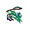 7pb8C 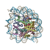 7piiC 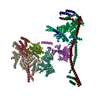 7pknC 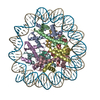 7r5rC 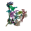 7r5sC 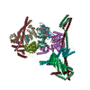 7r5vC 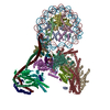 7ywxC 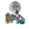 7yyhC  6ypcS S: Starting model for refinement C: citing same article ( |
|---|---|
| Similar structure data |


-Links
 PDBj
PDBj


- Assembly
| Deposited unit | 
| ||||||||
|---|---|---|---|---|---|---|---|---|---|
| 1 |
| ||||||||
| Unit cell |
|
-Components
| #1: Protein/peptide | Mass: 5726.729 Da / Num. of mol.: 1 Source method: isolated from a genetically manipulated source Source: (gene. exp.) Homo sapiens (human) / Gene: CENPH, ICEN35 / Production host:  |
|---|---|
| #2: Protein | Mass: 25832.582 Da / Num. of mol.: 1 Source method: isolated from a genetically manipulated source Source: (gene. exp.) Homo sapiens (human) / Gene: CENPI, FSHPRH1, ICEN19, LRPR1 / Production host: |
| #3: Protein | Mass: 12496.531 Da / Num. of mol.: 1 Source method: isolated from a genetically manipulated source Source: (gene. exp.) Homo sapiens (human) / Gene: CENPK, ICEN37, FKSG14 / Production host: |
-Experimental details
-Experiment
| Experiment | Method: X-RAY DIFFRACTION / Number of used crystals: 1 |
|---|
- Sample preparation
| Crystal | Density Matthews: 2.51 Å3/Da / Density % sol: 51.02 % |
|---|---|
| Crystal grow | Temperature: 277 K / Method: vapor diffusion, sitting drop / Details: 10% PEG 8k, 100 mM imidazole pH 8 |
-Data collection
| Diffraction | Mean temperature: 100 K / Serial crystal experiment: N |
|---|---|
| Diffraction source | Source: SYNCHROTRON / Site: Diamond / Beamline: I24 / Wavelength: 0.9795 Å |
| Detector | Type: DECTRIS PILATUS 6M / Detector: PIXEL / Date: Jan 26, 2021 |
| Radiation | Protocol: SINGLE WAVELENGTH / Monochromatic (M) / Laue (L): M / Scattering type: x-ray |
| Radiation wavelength | Wavelength: 0.9795 Å / Relative weight: 1 |
| Reflection | Resolution: 2.49→53.5 Å / Num. obs: 11771 / % possible obs: 99.5 % / Redundancy: 3.7 % / CC1/2: 0.98 / Rmerge(I) obs: 0.13 / Net I/σ(I): 8.5 |
| Reflection shell | Resolution: 2.49→2.6 Å / Rmerge(I) obs: 0.7 / Num. unique obs: 11771 / CC1/2: 0.3 |
- Processing
| Software |
| ||||||||||||||||||||||||||||||||||||||||
|---|---|---|---|---|---|---|---|---|---|---|---|---|---|---|---|---|---|---|---|---|---|---|---|---|---|---|---|---|---|---|---|---|---|---|---|---|---|---|---|---|---|
| Refinement | Method to determine structure: MOLECULAR REPLACEMENT Starting model: 6YPC Resolution: 2.49→47.37 Å / SU ML: 0.38 / Cross valid method: THROUGHOUT / σ(F): 1.34 / Phase error: 31.52 / Stereochemistry target values: ML
| ||||||||||||||||||||||||||||||||||||||||
| Solvent computation | Shrinkage radii: 0.9 Å / VDW probe radii: 1.11 Å / Solvent model: FLAT BULK SOLVENT MODEL | ||||||||||||||||||||||||||||||||||||||||
| Displacement parameters | Biso max: 143.83 Å2 / Biso mean: 50.5043 Å2 / Biso min: 15.03 Å2 | ||||||||||||||||||||||||||||||||||||||||
| Refinement step | Cycle: final / Resolution: 2.49→47.37 Å
| ||||||||||||||||||||||||||||||||||||||||
| LS refinement shell | Refine-ID: X-RAY DIFFRACTION / Rfactor Rfree error: 0 / Total num. of bins used: 4
| ||||||||||||||||||||||||||||||||||||||||
| Refinement TLS params. | Method: refined / Origin x: -19.2463 Å / Origin y: -6.7088 Å / Origin z: 21.7924 Å
| ||||||||||||||||||||||||||||||||||||||||
| Refinement TLS group |
|
