 Movie
Movie Controller


+ Open data

- Basic information
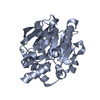
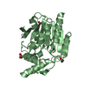
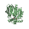
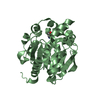
| Entry | Database: PDB / ID: 7p0y | |||||||||
|---|---|---|---|---|---|---|---|---|---|---|
| Title | Crystal Structure of mtbMGL K74A (Substrate Analog Complex) | |||||||||
 Components Components | Monoacylglycerol lipase | |||||||||
 Keywords Keywords | HYDROLASE / Mycobacterium tuberculosis / monoacylglycerol lipase | |||||||||
| Function / homology |  Function and homology information Function and homology informationglycerolipid catabolic process / acylglycerol lipase / lipase activity / monoacylglycerol lipase activity / peptidoglycan-based cell wall / symbiont-mediated activation of host apoptosis / extracellular region / membrane / plasma membrane Similarity search - Function | |||||||||
| Biological species |   Mycobacterium tuberculosis (bacteria) Mycobacterium tuberculosis (bacteria) | |||||||||
| Method |  X-RAY DIFFRACTION / SYNCHROTRON / MOLECULAR REPLACEMENT / Resolution: 2.25 Å X-RAY DIFFRACTION / SYNCHROTRON / MOLECULAR REPLACEMENT / Resolution: 2.25 Å | |||||||||
 Authors Authors | Grininger, C. / Aschauer, P. / Pavkov-Keller, T. / Oberer, M. | |||||||||
| Funding support |  Austria, 2items Austria, 2items
| |||||||||
 Citation Citation | Journal: Biomolecules / Year: 2021 Title: Structural Changes in the Cap of Rv0183/mtbMGL Modulate the Shape of the Binding Pocket. Authors: Grininger, C. / Leypold, M. / Aschauer, P. / Pavkov-Keller, T. / Riegler-Berket, L. / Breinbauer, R. / Oberer, M. | |||||||||
| History |
|
- Structure visualization
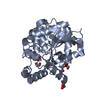
| Structure viewer | Molecule: MolmilJmol/JSmol |
|---|
- Downloads & links
-Download
| PDBx/mmCIF format | 7p0y.cif.gz | 132 KB | Display | PDBx/mmCIF format |
|---|---|---|---|---|
| PDB format | pdb7p0y.ent.gz | 93.1 KB | Display | PDB format |
| PDBx/mmJSON format | 7p0y.json.gz | Tree view | PDBx/mmJSON format | |
| Others |  Other downloads Other downloads |
-Validation report
| Arichive directory | https://data.pdbj.org/pub/pdb/validation_reports/p0/7p0yftp://data.pdbj.org/pub/pdb/validation_reports/p0/7p0y | HTTPS FTP |
|---|
-Related structure data
| Related structure data |  7ozmC 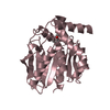 6eicS C: citing same article ( S: Starting model for refinement |
|---|---|
| Similar structure data |


-Links
 PDBj
PDBj

- Assembly
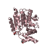
| Deposited unit | 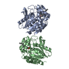
| ||||||||||||
|---|---|---|---|---|---|---|---|---|---|---|---|---|---|
| 1 | 
| ||||||||||||
| 2 | 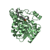
| ||||||||||||
| Unit cell |
|
-Components
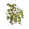
| #1: Protein | Mass: 30643.883 Da / Num. of mol.: 2 Source method: isolated from a genetically manipulated source Source: (gene. exp.) Mycobacterium tuberculosis (strain ATCC 25618 / H37Rv) (bacteria)Strain: ATCC 25618 / H37Rv / Gene: Rv0183, LH57_01015 / Production host: #2: Chemical | 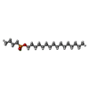  Mass: 364.518 Da / Num. of mol.: 2 / Source method: obtained synthetically / Formula: C20H42FO2P / Feature type: SUBJECT OF INVESTIGATION Mass: 364.518 Da / Num. of mol.: 2 / Source method: obtained synthetically / Formula: C20H42FO2P / Feature type: SUBJECT OF INVESTIGATION#3: Water | ChemComp-HOH / |  Mass: 18.015 Da / Num. of mol.: 141 / Source method: isolated from a natural source / Formula: H2O Mass: 18.015 Da / Num. of mol.: 141 / Source method: isolated from a natural source / Formula: H2OHas ligand of interest | Y | Has protein modification | Y | |
|---|
-Experimental details
-Experiment
| Experiment | Method: X-RAY DIFFRACTION / Number of used crystals: 1 |
|---|
- Sample preparation
| Crystal | Density Matthews: 2.36 Å3/Da / Density % sol: 47.88 % |
|---|---|
| Crystal grow | Temperature: 293 K / Method: vapor diffusion, sitting drop / pH: 8.5 Details: 30 % PEG300, 125 mM Calcium acetate, 100 mM Tris-HCl pH 8.5, 10 mg/ml protein |
-Data collection
| Diffraction | Mean temperature: 100 K / Serial crystal experiment: N |
|---|---|
| Diffraction source | Source: SYNCHROTRON / Site: PETRA III, DESY  / Beamline: P11 / Wavelength: 1.0332 Å / Beamline: P11 / Wavelength: 1.0332 Å |
| Detector | Type: DECTRIS PILATUS 6M / Detector: PIXEL / Date: Nov 10, 2019 |
| Radiation | Protocol: SINGLE WAVELENGTH / Monochromatic (M) / Laue (L): M / Scattering type: x-ray |
| Radiation wavelength | Wavelength: 1.0332 Å / Relative weight: 1 |
| Reflection | Resolution: 2.25→40.78 Å / Num. obs: 28192 / % possible obs: 99.65 % / Redundancy: 5.2 % / Biso Wilson estimate: 36.18 Å2 / CC1/2: 0.99 / CC star: 0.998 / Rmerge(I) obs: 0.149 / Rpim(I) all: 0.071 / Rrim(I) all: 0.165 / Net I/σ(I): 20.99 |
| Reflection shell | Resolution: 2.25→2.33 Å / Redundancy: 5.1 % / Rmerge(I) obs: 0.72 / Mean I/σ(I) obs: 1.19 / Num. unique obs: 2766 / CC1/2: 0.77 / CC star: 0.933 / Rpim(I) all: 0.35 / Rrim(I) all: 0.802 / % possible all: 99.5 |
- Processing
| Software |
| ||||||||||||||||||||||||||||||||||||||||||||||||||||||||||||||||||||||||||||||||||||||||||||||||||||||||||||||||||||||||||||||||||||||||||||||||||||||||||||||||||||||||||||||||||||||||||||||||||||
|---|---|---|---|---|---|---|---|---|---|---|---|---|---|---|---|---|---|---|---|---|---|---|---|---|---|---|---|---|---|---|---|---|---|---|---|---|---|---|---|---|---|---|---|---|---|---|---|---|---|---|---|---|---|---|---|---|---|---|---|---|---|---|---|---|---|---|---|---|---|---|---|---|---|---|---|---|---|---|---|---|---|---|---|---|---|---|---|---|---|---|---|---|---|---|---|---|---|---|---|---|---|---|---|---|---|---|---|---|---|---|---|---|---|---|---|---|---|---|---|---|---|---|---|---|---|---|---|---|---|---|---|---|---|---|---|---|---|---|---|---|---|---|---|---|---|---|---|---|---|---|---|---|---|---|---|---|---|---|---|---|---|---|---|---|---|---|---|---|---|---|---|---|---|---|---|---|---|---|---|---|---|---|---|---|---|---|---|---|---|---|---|---|---|---|---|---|---|
| Refinement | Method to determine structure: MOLECULAR REPLACEMENT Starting model: 6EIC Resolution: 2.25→40.78 Å / SU ML: 0.3809 / Cross valid method: FREE R-VALUE / σ(F): 1.34 / Phase error: 34.8137 Stereochemistry target values: GeoStd + Monomer Library + CDL v1.2
| ||||||||||||||||||||||||||||||||||||||||||||||||||||||||||||||||||||||||||||||||||||||||||||||||||||||||||||||||||||||||||||||||||||||||||||||||||||||||||||||||||||||||||||||||||||||||||||||||||||
| Solvent computation | Shrinkage radii: 0.9 Å / VDW probe radii: 1.11 Å / Solvent model: FLAT BULK SOLVENT MODEL | ||||||||||||||||||||||||||||||||||||||||||||||||||||||||||||||||||||||||||||||||||||||||||||||||||||||||||||||||||||||||||||||||||||||||||||||||||||||||||||||||||||||||||||||||||||||||||||||||||||
| Displacement parameters | Biso mean: 45.6 Å2 | ||||||||||||||||||||||||||||||||||||||||||||||||||||||||||||||||||||||||||||||||||||||||||||||||||||||||||||||||||||||||||||||||||||||||||||||||||||||||||||||||||||||||||||||||||||||||||||||||||||
| Refinement step | Cycle: LAST / Resolution: 2.25→40.78 Å
| ||||||||||||||||||||||||||||||||||||||||||||||||||||||||||||||||||||||||||||||||||||||||||||||||||||||||||||||||||||||||||||||||||||||||||||||||||||||||||||||||||||||||||||||||||||||||||||||||||||
| Refine LS restraints |
| ||||||||||||||||||||||||||||||||||||||||||||||||||||||||||||||||||||||||||||||||||||||||||||||||||||||||||||||||||||||||||||||||||||||||||||||||||||||||||||||||||||||||||||||||||||||||||||||||||||
| LS refinement shell |
|
