 Movie
Movie Controller

[English] 日本語
 Yorodumi
- PDB-7p0l: Crystal structure of S.pombe Mdb1 BRCT domains in complex with a ... -
+ Open data

- Basic information
| Entry | Database: PDB / ID: 7p0l | |||||||||
|---|---|---|---|---|---|---|---|---|---|---|
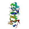
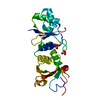
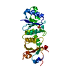
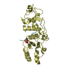
| Title | Crystal structure of S.pombe Mdb1 BRCT domains in complex with a H2A phosphopeptide | |||||||||
 Components Components |
| |||||||||
 Keywords Keywords | SIGNALING PROTEIN / DNA damage response | |||||||||
| Function / homology |  Function and homology information Function and homology informationCondensation of Prophase Chromosomes / : / : / Assembly of the ORC complex at the origin of replication / Oxidative Stress Induced Senescence / RMTs methylate histone arginines / Ub-specific processing proteases / HDACs deacetylate histones / Estrogen-dependent gene expression / RNA Polymerase I Promoter Escape ...Condensation of Prophase Chromosomes / : / : / Assembly of the ORC complex at the origin of replication / Oxidative Stress Induced Senescence / RMTs methylate histone arginines / Ub-specific processing proteases / HDACs deacetylate histones / Estrogen-dependent gene expression / RNA Polymerase I Promoter Escape / Recruitment and ATM-mediated phosphorylation of repair and signaling proteins at DNA double strand breaks / Transcriptional regulation by small RNAs / mating-type region heterochromatin / mitotic DNA damage checkpoint signaling / chromosome, subtelomeric region / mitotic spindle midzone / homologous chromosome segregation / rDNA heterochromatin / mitotic chromosome condensation / chromatin-protein adaptor activity / mitotic G2 DNA damage checkpoint signaling / pericentric heterochromatin / structural constituent of chromatin / heterochromatin formation / nucleosome / double-strand break repair / site of double-strand break / protein heterodimerization activity / DNA binding / nucleus / cytoplasm Similarity search - Function | |||||||||
| Biological species |  | |||||||||
| Method |  X-RAY DIFFRACTION / SYNCHROTRON / MOLECULAR REPLACEMENT / Resolution: 1.97 Å X-RAY DIFFRACTION / SYNCHROTRON / MOLECULAR REPLACEMENT / Resolution: 1.97 Å | |||||||||
 Authors Authors | Day, M. / Oliver, A.W. / Pearl, L.H. | |||||||||
| Funding support |  United Kingdom, 2items United Kingdom, 2items
| |||||||||
 Citation Citation | Journal: DNA Repair (Amst) / Year: 2021 Title: Phosphorylation-dependent assembly of DNA damage response systems and the central roles of TOPBP1. Authors: Day, M. / Oliver, A.W. / Pearl, L.H. #1: Journal: Acta Crystallogr., Sect. D: Biol. Crystallogr. / Year: 2012Title: Towards automated crystallographic structure refinement with phenix.refine. Authors: Afonine, P.V. | |||||||||
| History |
|
- Structure visualization
| Structure viewer | Molecule: MolmilJmol/JSmol |
|---|
- Downloads & links
-Download
| PDBx/mmCIF format | 7p0l.cif.gz | 198.6 KB | Display | PDBx/mmCIF format |
|---|---|---|---|---|
| PDB format | pdb7p0l.ent.gz | 129 KB | Display | PDB format |
| PDBx/mmJSON format | 7p0l.json.gz | Tree view | PDBx/mmJSON format | |
| Others |  Other downloads Other downloads |
-Validation report
| Arichive directory | https://data.pdbj.org/pub/pdb/validation_reports/p0/7p0lftp://data.pdbj.org/pub/pdb/validation_reports/p0/7p0l | HTTPS FTP |
|---|
-Related structure data
| Related structure data |  7p0jSC S: Starting model for refinement C: citing same article ( |
|---|---|
| Similar structure data |


-Links
 PDBj
PDBj


- Assembly
| Deposited unit | 
| |||||||||||||||||||||
|---|---|---|---|---|---|---|---|---|---|---|---|---|---|---|---|---|---|---|---|---|---|---|
| 1 | 
| |||||||||||||||||||||
| 2 | 
| |||||||||||||||||||||
| Unit cell |
| |||||||||||||||||||||
| Noncrystallographic symmetry (NCS) | NCS domain:
NCS domain segments: Ens-ID: 1 / Beg auth comp-ID: THR / Beg label comp-ID: THR / End auth comp-ID: ASP / End label comp-ID: ASP / Auth seq-ID: 387 - 581 / Label seq-ID: 5 - 199
|
-Components
| #1: Protein | Mass: 22696.945 Da / Num. of mol.: 2 Source method: isolated from a genetically manipulated source Source: (gene. exp.) Strain: 972 / ATCC 24843 / Gene: mdb1, SPAC2E11.14, SPACUNK4.14 / Production host:  #2: Protein/peptide | Mass: 1241.246 Da / Num. of mol.: 2 / Source method: obtained synthetically Source: (synth.) References: UniProt: P04910 #3: Water | ChemComp-HOH / |  Mass: 18.015 Da / Num. of mol.: 159 / Source method: isolated from a natural source / Formula: H2O Mass: 18.015 Da / Num. of mol.: 159 / Source method: isolated from a natural source / Formula: H2OHas ligand of interest | Y | Has protein modification | Y | |
|---|
-Experimental details
-Experiment
| Experiment | Method: X-RAY DIFFRACTION / Number of used crystals: 1 |
|---|
- Sample preparation
| Crystal | Density Matthews: 1.73 Å3/Da / Density % sol: 28.93 % |
|---|---|
| Crystal grow | Temperature: 277 K / Method: vapor diffusion, sitting drop Details: 200mM Magnesium Chloride 100mM TRIS pH 8.0 30% (w/v) PEG 4000 |
-Data collection
| Diffraction | Mean temperature: 100 K / Serial crystal experiment: N |
|---|---|
| Diffraction source | Source: SYNCHROTRON / Site: Diamond / Beamline: I03 / Wavelength: 0.97957 Å |
| Detector | Type: DECTRIS PILATUS 6M / Detector: PIXEL / Date: Aug 11, 2014 |
| Radiation | Protocol: SINGLE WAVELENGTH / Monochromatic (M) / Laue (L): M / Scattering type: x-ray |
| Radiation wavelength | Wavelength: 0.97957 Å / Relative weight: 1 |
| Reflection | Resolution: 1.97→48.12 Å / Num. obs: 23405 / % possible obs: 98.7 % / Redundancy: 3.2 % / Biso Wilson estimate: 35.94 Å2 / Rpim(I) all: 0.044 / Net I/σ(I): 1.087 |
| Reflection shell | Resolution: 1.97→2.02 Å / Num. unique obs: 1720 / Rpim(I) all: 0.383 |
- Processing
| Software |
| |||||||||||||||||||||||||||||||||||||||||||||||||||||||||||||||
|---|---|---|---|---|---|---|---|---|---|---|---|---|---|---|---|---|---|---|---|---|---|---|---|---|---|---|---|---|---|---|---|---|---|---|---|---|---|---|---|---|---|---|---|---|---|---|---|---|---|---|---|---|---|---|---|---|---|---|---|---|---|---|---|---|
| Refinement | Method to determine structure: MOLECULAR REPLACEMENT Starting model: 7P0J Resolution: 1.97→48.12 Å / SU ML: 0.2648 / Cross valid method: FREE R-VALUE / σ(F): 1.34 / Phase error: 28.0307 Stereochemistry target values: GeoStd + Monomer Library + CDL v1.2
| |||||||||||||||||||||||||||||||||||||||||||||||||||||||||||||||
| Solvent computation | Shrinkage radii: 0.9 Å / VDW probe radii: 1.11 Å / Solvent model: FLAT BULK SOLVENT MODEL | |||||||||||||||||||||||||||||||||||||||||||||||||||||||||||||||
| Displacement parameters | Biso mean: 48.47 Å2 | |||||||||||||||||||||||||||||||||||||||||||||||||||||||||||||||
| Refinement step | Cycle: LAST / Resolution: 1.97→48.12 Å
| |||||||||||||||||||||||||||||||||||||||||||||||||||||||||||||||
| Refine LS restraints |
| |||||||||||||||||||||||||||||||||||||||||||||||||||||||||||||||
| LS refinement shell |
|
