 Movie
Movie Controller


+ Open data

- Basic information
| Entry | Database: PDB / ID: 7oxi | |||||||||
|---|---|---|---|---|---|---|---|---|---|---|
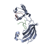
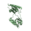
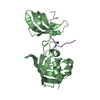
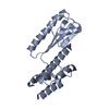
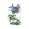
| Title | ttSlyD with W4A pseudo-wild-type S2 peptide | |||||||||
 Components Components |
| |||||||||
 Keywords Keywords | ISOMERASE / FKBP / chaperone | |||||||||
| Function / homology |  Function and homology information Function and homology informationpeptidylprolyl isomerase / peptidyl-prolyl cis-trans isomerase activity / protein refolding / ribosomal small subunit assembly / cytosolic small ribosomal subunit / cytoplasmic translation / structural constituent of ribosome / zinc ion binding / metal ion binding / cytoplasm Similarity search - Function | |||||||||
| Biological species |   Thermus thermophilus (bacteria) Thermus thermophilus (bacteria) | |||||||||
| Method |  X-RAY DIFFRACTION / SYNCHROTRON / MOLECULAR REPLACEMENT / Resolution: 2.6 Å X-RAY DIFFRACTION / SYNCHROTRON / MOLECULAR REPLACEMENT / Resolution: 2.6 Å | |||||||||
 Authors Authors | Pazicky, S. / Lei, J. / Loew, C. | |||||||||
| Funding support |  Germany, 2items Germany, 2items
| |||||||||
 Citation Citation | Journal: Cell.Mol.Life Sci. / Year: 2022 Title: Impact of distant peptide substrate residues on enzymatic activity of SlyD. Authors: Pazicky, S. / Werle, A.A. / Lei, J. / Low, C. / Weininger, U. | |||||||||
| History |
|
- Structure visualization
| Structure viewer | Molecule: MolmilJmol/JSmol |
|---|
- Downloads & links
-Download
| PDBx/mmCIF format | 7oxi.cif.gz | 98 KB | Display | PDBx/mmCIF format |
|---|---|---|---|---|
| PDB format | pdb7oxi.ent.gz | 64 KB | Display | PDB format |
| PDBx/mmJSON format | 7oxi.json.gz | Tree view | PDBx/mmJSON format | |
| Others |  Other downloads Other downloads |
-Validation report
| Arichive directory | https://data.pdbj.org/pub/pdb/validation_reports/ox/7oxiftp://data.pdbj.org/pub/pdb/validation_reports/ox/7oxi | HTTPS FTP |
|---|
-Related structure data
| Related structure data |  7oxgC  7oxhC  7oxjC  7oxkC  3cgmS S: Starting model for refinement C: citing same article ( |
|---|---|
| Similar structure data |


-Links
 PDBj
PDBj


- Assembly
| Deposited unit | 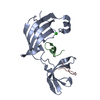
| ||||||||||
|---|---|---|---|---|---|---|---|---|---|---|---|
| 1 |
| ||||||||||
| Unit cell |
|
-Components
| #1: Protein | Mass: 17400.234 Da / Num. of mol.: 1 Source method: isolated from a genetically manipulated source Source: (gene. exp.) Thermus thermophilus (strain ATCC 27634 / DSM 579 / HB8) (bacteria)Strain: ATCC 27634 / DSM 579 / HB8 / Gene: TTHA0346 / Production host: | ||||||
|---|---|---|---|---|---|---|---|
| #2: Protein/peptide | Mass: 1659.948 Da / Num. of mol.: 2 / Mutation: W23E, P25A, K28L, I30A / Source method: obtained synthetically / Source: (synth.) #3: Chemical | ChemComp-CL / |   Mass: 35.453 Da / Num. of mol.: 1 / Source method: obtained synthetically / Formula: Cl Mass: 35.453 Da / Num. of mol.: 1 / Source method: obtained synthetically / Formula: Cl#4: Water | ChemComp-HOH / |  Mass: 18.015 Da / Num. of mol.: 4 / Source method: isolated from a natural source / Formula: H2O Mass: 18.015 Da / Num. of mol.: 4 / Source method: isolated from a natural source / Formula: H2OHas ligand of interest | N | |
-Experimental details
-Experiment
| Experiment | Method: X-RAY DIFFRACTION / Number of used crystals: 1 |
|---|
- Sample preparation
| Crystal | Density Matthews: 4.09 Å3/Da / Density % sol: 69.96 % |
|---|---|
| Crystal grow | Temperature: 292.15 K / Method: counter-diffusion / pH: 6.5 / Details: sodium cacodylate, MPD, PEG8000 |
-Data collection
| Diffraction | Mean temperature: 100 K / Serial crystal experiment: N |
|---|---|
| Diffraction source | Source: SYNCHROTRON / Site: PETRA III, EMBL c/o DESY / Beamline: P13 (MX1) / Wavelength: 1.0332 Å |
| Detector | Type: DECTRIS PILATUS 6M / Detector: PIXEL / Date: Mar 14, 2019 |
| Radiation | Protocol: SINGLE WAVELENGTH / Monochromatic (M) / Laue (L): M / Scattering type: x-ray |
| Radiation wavelength | Wavelength: 1.0332 Å / Relative weight: 1 |
| Reflection | Resolution: 2.6→41.86 Å / Num. obs: 10215 / % possible obs: 99.9 % / Redundancy: 18.5 % / Biso Wilson estimate: 95.41 Å2 / CC1/2: 0.999 / Rmerge(I) obs: 0.0567 / Rpim(I) all: 0.0139 / Rrim(I) all: 0.05847 / Net I/σ(I): 30.51 |
| Reflection shell | Resolution: 2.6→2.7 Å / Redundancy: 17.9 % / Rmerge(I) obs: 2.105 / Mean I/σ(I) obs: 1.43 / Num. unique obs: 988 / CC1/2: 0.704 / Rpim(I) all: 0.5057 / Rrim(I) all: 2.166 / % possible all: 99.4 |
- Processing
| Software |
| ||||||||||||||||||||||||||||||||||||||||
|---|---|---|---|---|---|---|---|---|---|---|---|---|---|---|---|---|---|---|---|---|---|---|---|---|---|---|---|---|---|---|---|---|---|---|---|---|---|---|---|---|---|
| Refinement | Method to determine structure: MOLECULAR REPLACEMENT Starting model: 3cgm Resolution: 2.6→41.86 Å / SU ML: 0.3141 / Cross valid method: FREE R-VALUE / σ(F): 1.34 / Phase error: 37.9729 Stereochemistry target values: GeoStd + Monomer Library + CDL v1.2
| ||||||||||||||||||||||||||||||||||||||||
| Solvent computation | Shrinkage radii: 0.9 Å / VDW probe radii: 1.11 Å / Solvent model: FLAT BULK SOLVENT MODEL | ||||||||||||||||||||||||||||||||||||||||
| Displacement parameters | Biso mean: 119.85 Å2 | ||||||||||||||||||||||||||||||||||||||||
| Refinement step | Cycle: LAST / Resolution: 2.6→41.86 Å
| ||||||||||||||||||||||||||||||||||||||||
| Refine LS restraints |
| ||||||||||||||||||||||||||||||||||||||||
| LS refinement shell |
| ||||||||||||||||||||||||||||||||||||||||
| Refinement TLS params. | Method: refined / Origin x: 16.4402406388 Å / Origin y: 57.0743859762 Å / Origin z: 38.7897743263 Å
| ||||||||||||||||||||||||||||||||||||||||
| Refinement TLS group | Selection details: all |
