 Movie
Movie Controller

[English] 日本語
 Yorodumi
- PDB-3uqb: Crystal structure of a SMT Fusion PEPTIDYL-PROLYL CIS-TRANS ISOME... -
+ Open data

- Basic information
| Entry | Database: PDB / ID: 3uqb | ||||||
|---|---|---|---|---|---|---|---|
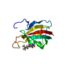
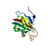
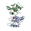
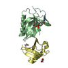
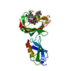
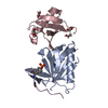
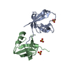
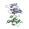
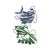
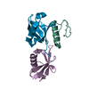
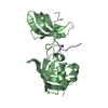
| Title | Crystal structure of a SMT Fusion PEPTIDYL-PROLYL CIS-TRANS ISOMERASE with surface mutation D44G from Burkholderia pseudomallei complexed with FK506 | ||||||
 Components Components | Ubiquitin-like protein SMT3, Peptidyl-prolyl cis-trans isomerase | ||||||
 Keywords Keywords | Isomerase / protein binding / SSGCID / Structural Genomics / Seattle Structural Genomics Center for Infectious Disease | ||||||
| Function / homology |  Function and homology information Function and homology informationSUMO is conjugated to E1 (UBA2:SAE1) / SUMOylation of nuclear envelope proteins / SUMO is transferred from E1 to E2 (UBE2I, UBC9) / SUMO is proteolytically processed / SUMOylation of transcription factors / Postmitotic nuclear pore complex (NPC) reformation / SUMOylation of transcription cofactors / septin ring / SUMOylation of DNA damage response and repair proteins / Transcriptional and post-translational regulation of MITF-M expression and activity ...SUMO is conjugated to E1 (UBA2:SAE1) / SUMOylation of nuclear envelope proteins / SUMO is transferred from E1 to E2 (UBE2I, UBC9) / SUMO is proteolytically processed / SUMOylation of transcription factors / Postmitotic nuclear pore complex (NPC) reformation / SUMOylation of transcription cofactors / septin ring / SUMOylation of DNA damage response and repair proteins / Transcriptional and post-translational regulation of MITF-M expression and activity / SUMOylation of DNA replication proteins / SUMOylation of SUMOylation proteins / Recruitment and ATM-mediated phosphorylation of repair and signaling proteins at DNA double strand breaks / SUMOylation of RNA binding proteins / SUMOylation of chromatin organization proteins / ubiquitin-like protein ligase binding / protein sumoylation / condensed nuclear chromosome / peptidylprolyl isomerase / peptidyl-prolyl cis-trans isomerase activity / protein tag activity / metal ion binding / identical protein binding / nucleus Similarity search - Function | ||||||
| Biological species |   Burkholderia pseudomallei (bacteria) Burkholderia pseudomallei (bacteria) | ||||||
| Method |  X-RAY DIFFRACTION / SYNCHROTRON / molecular replacement / Resolution: 1.9 Å X-RAY DIFFRACTION / SYNCHROTRON / molecular replacement / Resolution: 1.9 Å | ||||||
 Authors Authors | Seattle Structural Genomics Center for Infectious Disease (SSGCID) | ||||||
 Citation Citation | Journal: Antimicrob.Agents Chemother. / Year: 2014 Title: A structural biology approach enables the development of antimicrobials targeting bacterial immunophilins. Authors: Begley, D.W. / Fox, D. / Jenner, D. / Juli, C. / Pierce, P.G. / Abendroth, J. / Muruthi, M. / Safford, K. / Anderson, V. / Atkins, K. / Barnes, S.R. / Moen, S.O. / Raymond, A.C. / Stacy, R. ...Authors: Begley, D.W. / Fox, D. / Jenner, D. / Juli, C. / Pierce, P.G. / Abendroth, J. / Muruthi, M. / Safford, K. / Anderson, V. / Atkins, K. / Barnes, S.R. / Moen, S.O. / Raymond, A.C. / Stacy, R. / Myler, P.J. / Staker, B.L. / Harmer, N.J. / Norville, I.H. / Holzgrabe, U. / Sarkar-Tyson, M. / Edwards, T.E. / Lorimer, D.D. | ||||||
| History |
|
- Structure visualization
| Structure viewer | Molecule: MolmilJmol/JSmol |
|---|
- Downloads & links
-Download
| PDBx/mmCIF format | 3uqb.cif.gz | 92.2 KB | Display | PDBx/mmCIF format |
|---|---|---|---|---|
| PDB format | pdb3uqb.ent.gz | 68.2 KB | Display | PDB format |
| PDBx/mmJSON format | 3uqb.json.gz | Tree view | PDBx/mmJSON format | |
| Others |  Other downloads Other downloads |
-Validation report
| Arichive directory | https://data.pdbj.org/pub/pdb/validation_reports/uq/3uqbftp://data.pdbj.org/pub/pdb/validation_reports/uq/3uqb | HTTPS FTP |
|---|
-Related structure data
| Related structure data | 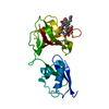 3uf8SC 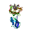 3uqaC  3vawC 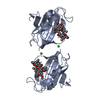 4dz2C 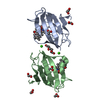 4dz3C 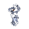 4fn2C 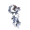 4g50C 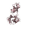 4ggqC 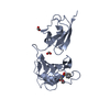 4givC S: Starting model for refinement C: citing same article ( |
|---|---|
| Similar structure data | |
| Other databases |


-Links
 PDBj
PDBj


- Assembly
| Deposited unit | 
| ||||||||
|---|---|---|---|---|---|---|---|---|---|
| 1 |
| ||||||||
| Unit cell |
|
-Components
| #1: Protein | Mass: 22943.793 Da / Num. of mol.: 1 / Fragment: Q12306 residues 13-98, Q3JK38 residues 2-113 / Mutation: D44G Source method: isolated from a genetically manipulated source Details: Fusion Protein Source: (gene. exp.) Burkholderia pseudomallei (bacteria)Strain: 1710b, S288c / Gene: BURPS1710b_A0907, SMT3, YDR510W, D9719.15 / Plasmid: pET28-HisSMT / Production host: References: UniProt: Q12306, UniProt: Q3JK38, peptidylprolyl isomerase |
|---|---|
| #2: Chemical | ChemComp-FK5 /   Mass: 804.018 Da / Num. of mol.: 1 / Source method: obtained synthetically / Formula: C44H69NO12 / Comment: medication*YM Mass: 804.018 Da / Num. of mol.: 1 / Source method: obtained synthetically / Formula: C44H69NO12 / Comment: medication*YM |
| #3: Water | ChemComp-HOH /  Mass: 18.015 Da / Num. of mol.: 163 / Source method: isolated from a natural source / Formula: H2O Mass: 18.015 Da / Num. of mol.: 163 / Source method: isolated from a natural source / Formula: H2O |
-Experimental details
-Experiment
| Experiment | Method: X-RAY DIFFRACTION / Number of used crystals: 1 |
|---|
- Sample preparation
| Crystal | Density Matthews: 1.99 Å3/Da / Density % sol: 38.25 % |
|---|---|
| Crystal grow | Temperature: 290 K / Method: vapor diffusion, sitting drop / pH: 9 Details: Internal tracking number 226421. PACT well B6. 0.1M MIB Buffer pH 9.0, 25.0% w/v PEG1500, 30% PEG400 Cryo. BUPSA.00130.A.D214 PD00190/6 23.7mg/ml, vapor diffusion, sitting drop, temperature ...Details: Internal tracking number 226421. PACT well B6. 0.1M MIB Buffer pH 9.0, 25.0% w/v PEG1500, 30% PEG400 Cryo. BUPSA.00130.A.D214 PD00190/6 23.7mg/ml, vapor diffusion, sitting drop, temperature 290K, VAPOR DIFFUSION, SITTING DROP |
-Data collection
| Diffraction | Mean temperature: 100 K | |||||||||||||||||||||||||||||||||||||||||||||||||||||||||||||||||||||||||||||||||||||||||||||||||||||||||||||||||||||||||||||||||||||||||||||||||||
|---|---|---|---|---|---|---|---|---|---|---|---|---|---|---|---|---|---|---|---|---|---|---|---|---|---|---|---|---|---|---|---|---|---|---|---|---|---|---|---|---|---|---|---|---|---|---|---|---|---|---|---|---|---|---|---|---|---|---|---|---|---|---|---|---|---|---|---|---|---|---|---|---|---|---|---|---|---|---|---|---|---|---|---|---|---|---|---|---|---|---|---|---|---|---|---|---|---|---|---|---|---|---|---|---|---|---|---|---|---|---|---|---|---|---|---|---|---|---|---|---|---|---|---|---|---|---|---|---|---|---|---|---|---|---|---|---|---|---|---|---|---|---|---|---|---|---|---|---|
| Diffraction source | Source: SYNCHROTRON / Site: ALS  / Beamline: 5.0.1 / Wavelength: 0.9774 Å / Beamline: 5.0.1 / Wavelength: 0.9774 Å | |||||||||||||||||||||||||||||||||||||||||||||||||||||||||||||||||||||||||||||||||||||||||||||||||||||||||||||||||||||||||||||||||||||||||||||||||||
| Detector | Type: ADSC QUANTUM 315r / Detector: CCD / Date: Oct 16, 2011 | |||||||||||||||||||||||||||||||||||||||||||||||||||||||||||||||||||||||||||||||||||||||||||||||||||||||||||||||||||||||||||||||||||||||||||||||||||
| Radiation | Monochromator: Si(220) Asymmetric cut single crystal / Protocol: SINGLE WAVELENGTH / Monochromatic (M) / Laue (L): M / Scattering type: x-ray | |||||||||||||||||||||||||||||||||||||||||||||||||||||||||||||||||||||||||||||||||||||||||||||||||||||||||||||||||||||||||||||||||||||||||||||||||||
| Radiation wavelength | Wavelength: 0.9774 Å / Relative weight: 1 | |||||||||||||||||||||||||||||||||||||||||||||||||||||||||||||||||||||||||||||||||||||||||||||||||||||||||||||||||||||||||||||||||||||||||||||||||||
| Reflection | Resolution: 1.9→38.5 Å / Num. all: 14561 / Num. obs: 14527 / % possible obs: 99.8 % / Observed criterion σ(F): -3 / Observed criterion σ(I): -3 / Redundancy: 4.02 % / Biso Wilson estimate: 24.794 Å2 / Rmerge(I) obs: 0.07 / Net I/σ(I): 16.55 | |||||||||||||||||||||||||||||||||||||||||||||||||||||||||||||||||||||||||||||||||||||||||||||||||||||||||||||||||||||||||||||||||||||||||||||||||||
| Reflection shell | Diffraction-ID: 1
|
-Phasing
| Phasing | Method: molecular replacement |
|---|
- Processing
| Software |
| ||||||||||||||||||||||||||||||||||||||||||||||||||||||||||||
|---|---|---|---|---|---|---|---|---|---|---|---|---|---|---|---|---|---|---|---|---|---|---|---|---|---|---|---|---|---|---|---|---|---|---|---|---|---|---|---|---|---|---|---|---|---|---|---|---|---|---|---|---|---|---|---|---|---|---|---|---|---|
| Refinement | Method to determine structure: molecular replacement Starting model: PDB entry 3UF8 Resolution: 1.9→19.25 Å / Cor.coef. Fo:Fc: 0.956 / Cor.coef. Fo:Fc free: 0.919 / SU B: 5.59 / SU ML: 0.094 / Cross valid method: THROUGHOUT / σ(F): 0 / σ(I): 2 / ESU R: 0.159 / ESU R Free: 0.154 / Stereochemistry target values: MAXIMUM LIKELIHOOD Details: U VALUES : WITH TLS ADDED HYDROGENS HAVE BEEN ADDED IN THE RIDING POSITIONS
| ||||||||||||||||||||||||||||||||||||||||||||||||||||||||||||
| Solvent computation | Ion probe radii: 0.8 Å / Shrinkage radii: 0.8 Å / VDW probe radii: 1.2 Å / Solvent model: MASK | ||||||||||||||||||||||||||||||||||||||||||||||||||||||||||||
| Displacement parameters | Biso mean: 19.783 Å2
| ||||||||||||||||||||||||||||||||||||||||||||||||||||||||||||
| Refinement step | Cycle: LAST / Resolution: 1.9→19.25 Å
| ||||||||||||||||||||||||||||||||||||||||||||||||||||||||||||
| Refine LS restraints |
| ||||||||||||||||||||||||||||||||||||||||||||||||||||||||||||
| LS refinement shell | Resolution: 1.9→1.949 Å / Total num. of bins used: 20
| ||||||||||||||||||||||||||||||||||||||||||||||||||||||||||||
| Refinement TLS params. | Method: refined / Origin x: 22.6918 Å / Origin y: 0.6483 Å / Origin z: 58.096 Å
|
